THE PRESENCE OF ANTIOXIDANTS IN THE HAIR INFUNDIBULUM IMPLICATIONS IN HAIR DISEASES SUCH AS ALOPECIA
1 MBA, 13442 SW 102 Lane Miami, Florida
33186, USA
|
|
|
ABSTRACT |
|
|
Anatomically,
as a rule the hair in mammals consists of an unseen follicle or root anchored
into the skin with a shaft or visible hair exiting exteriorly. As a note of
interest, the hair follicle has been described as a miniorgan having its own
cells division, metabolism, and aging stages Schneider et al. (2009). As previously stated, “metabolism entails electron transfers in
both plants (photosynthesis) and animals (cellular respiration) involving
movement of electrons from donor to acceptor along the electron transfer
chain thus inducing a current within each cell and from cell to cell” Embi et al. (2015), Scherlag et al. (2016). This continuity of energy transfer in living organisms is at the
very essence of life; and is ubiquitously present in all living matter and
the generator of Bioelectricity (a.k.a. electromagnetic radiation), the
protein enzyme catalase having an essential pivotal role in energy production
in the breakdown of toxic materials such as H2O2 into H2O and O2 molecules.
During the breakdown of O2 molecules energy is generated. When catalase is
depleted life ends and regional death occurs Embi (2018), Levin (2014). We could then theorize that if cell respiration ceases
throughout the entire organism (organs) death ensues Embi (2018). Only in living tissue is that elimination of toxic material such
as H2O2 has any relevance. |
|||
|
Received 11 April 2022 Accepted 16 May 2022 Published 31 May 2022 Corresponding Author Abraham
A. Embi, DOI 10.29121/granthaalayah.v10.i5.2022.4593 Funding: This research
received no specific grant from any funding agency in the public, commercial,
or not-for-profit sectors. Copyright: © 2022 The
Author(s). This work is licensed under a Creative Commons
Attribution 4.0 International License. With the
license CC-BY, authors retain the copyright, allowing anyone to download,
reuse, re-print, modify, distribute, and/or copy their contribution. The work
must be properly attributed to its author.
|
|||
|
Keywords: Exogenous Material Entering the
Hair/Shaft Skin Cavity, and The Presence of Antioxidants |
|||
1. INTRODUCTION
Now that we have established that antioxidants are the essence of life, is time to introduce In Vitro experiments inferring the presence of said antioxidants at the hair/shaft/skin junction and their possible implications. Prior In Vitro experiments by this author has shown the prevalence of bioelectrical crosstalk amongst human tissues, namely the hair and blood Embi (2018). The presence of antioxidants in the hair/shaft skin junction could be also classified as a facilitator of bioelectrical crosstalk between exogenous matter causing diseases such as mites and bacteria or chemicals entering the most proximal distal hair area at the skin/shaft junction or Infundibulum as shown in Figure 1, Figure 2, Figure 3 plus videos.
“The hair root is in the skin and extends down to the deeper layers of the skin. It is surrounded by the hair follicle (a sheath of skin and connective tissue) National Library of Medicine (2019).
The human hair anatomical image on paper is conventionally shown as an artist drawing; usually depicted as follows in Exhibit 1
EXHIBIT I
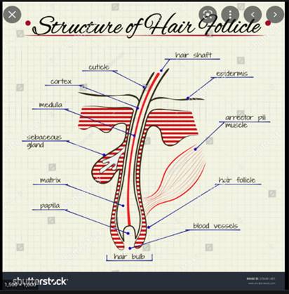
|
EXHIBIT 1 The human hair anatomical image |
2. VIDEO MICROSCOPY IMAGE
The Attracting Property of Hair Shaft/Skin Junction Environment
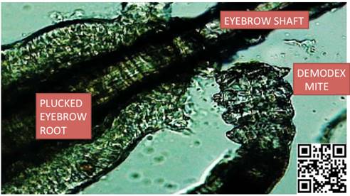
Unpublished Image showing skin parasite attracted to human eyebrow hair shaft/skin junction.
Figure 1
|
Figure 1 Skin Parasite Attracted to Human Eyebrow Hair Shaft/Skin Junction |
Figure 1 Incidental finding Microscopy image magnified x40. Skin parasite found (Mite) found while doing catalase research. Plucked one of my eyebrows, mounted in microscope and there it was living at the shaft/skin junction. For details link to: https://youtu.be/YBdJqhF9QI0
Or Scan QR Code in lower right corner of image.
3. DEMONSTRATION OF THE PRESENCE OF ANTIOXIDANTS IN THE HAIR INFUNDIBULUM
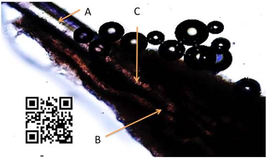
The attraction of H2O2 molecules in water being attracted into the hair infundibulum, them broken down by antioxidants in the infundibulum into H2O2 and O2 molecules. Figure below showing O2 molecules exiting hair.
Figure 2
|
Figure 2 O2 molecules exiting hair |
Figure 2 Demonstration of antioxidants breaking down H2O2 in infundibulum. Microphotograph of video. Plucked scalp hair immersed in water with H2O2 molecules. A= Hair Shaft B= Oxygen Bubbles from H2O2 decomposition by catalase C= Hair Follicle Infundibulum
For additional details link to https://youtu.be/09tYp348jKM. Or scan QR Code in left side of image.
Image reproduced from: Abraham Embi. The secondary role of UV light in swimmer’s melanoma genesis. Int J Mol Biol Open Access. 2018;3(3):121-123. DOI: 10.15406/ijmboa.2018.03.00064
Images below Figure 3 supplemental information obtained while researching the hair follicle bio magnetic imprint. See citation below.
4. HUMAN HAIR HAIR BIOELECTRIC IMPRINT
4.1. PLEASE NOTICE DIFFERENCE WHEN COMPARING WITH EXHIBIT I
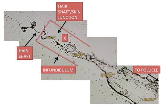
Figure 3
|
Figure 3 Scattered Bioelectrical Activity in Infundibulum |
Figure 3 Distal hair bioelectric magnetic imprint showing scattered bioelectrical activity in infundibulum. X: Notice scattered bioelectric activity in area surrounded by red lines (infundibulum). This activity is in support of the presence of antioxidants.
How to obtain a bioelectrical imprint of the hair is fully explained by linking to:
Embi AA. Introducing Gap in Hair Follicle Electromagnetism as Proposed Mechanism for The Presence of Bipolar Electrical Charges Inherent in The Human Hair Shaft. DOI 10.29121/granthaalayah.v9.i9.2021.4260
5. THE QUESTIONS ARISE:
1) Why are microbes and parasites attracted to the human hair shaft/skin junction? Are antioxidants attracting parasites?
2) Does the presence of a Biolectromagnetic environment is a factor in the attraction?
3) Is hair follicle inflammation as in alopecia caused by microorganisms reacting to antioxidants present in the infundibulum? After all some diseases such as esophageal reflux are caused by helicobacter pylori.
4) Further research is highly recommended!!
CONFLICT OF INTERESTS
None.
ACKNOWLEDGMENTS
None.
REFERENCES
Embi, A. A. (2018). Introducing Antioxidants As Essential For The Maintenance Of Tissue Life As Demonstrated In Human Hair Follicles. International Journal of Research -GRANTHAALAYAH, 6(7), 263-271. https://doi.org/10.29121/granthaalayah.v6.i7.2018.1305
Embi, A.A. (2018). Hair and blood endogenous low level biomagnetic fields cross-talk effects on fibrin inhibition and rouleaux formation. IJGR, 2018 6(11), 200-208. https://doi.org/10.29121/granthaalayah.v6.i11.2018.1118
Embi, A.A. Jacobson, J.I. Sahoo, K. Scherlag, B.J. (2015). Demonstration of Inherent Electromagnetic Energy Emanating from Isolated Human Hairs.
Embi, A.A.B.s. (2018). Compatibility Of Biomagnetic Profiles Found In Living Matter By Cross Species Demonstration. International Journal of Research - Granthaalayah, 6(8), 84-92. https://doi.org/10.29121/granthaalayah.v6.i8.2018.1264
Levin, M. (2014). Molecular bioelectricity : How endogenous voltage potentials control cell behavior and instruct pattern regulation in vivo. Molecular Biology of the Cell. 25 (24). https://doi.org/10.1091/mbc.e13-12-0708
National Library of Medicine (2019). What is the structure of hair and how does it grow ?
Scherlag, B.J. Sahoo, K. Embi, A.A. (2016). Novel and Simplified Method for Imaging the Electromagnetic Energy in Plant and Animal Tissues. Journal of Nanoscience and Nanoengineering, 2(1), 6-9.
Schneider, M.R. Schmidt-Ullrich. R. Paus, R. (2009). The hair follicle as a dynamic miniorgan. 19(3). https://doi.org/10.1016/j.cub.2008.12.005
 This work is licensed under a: Creative Commons Attribution 4.0 International License
© Granthaalayah 2014-2022. All Rights Reserved.