Original Article
Molecular and Cellular Evaluation of Anticancer Activities of Selected Indian Herbal Extracts: Focus on Curcuma longa, Withania somnifera, and Ocimum
|
Dr. Kaina
Bhonsle 1*, Dr. Haish Vyas 2, Dr. Riddhi Pradhan 3, Dr.
Alka Vyas 4 1 Faculty of Microbiology,
Samrat Vikramaditya Vishwavidhyalay, Ujjain, India 2 Principal, Madhav Science College, Ujjain,
India 3 Associate Professor, Microbiology R. D. Gardi
Medical College, Ujjain, India 4 Head of the Department Microbiology and Food
Technology, Samrat Vikramaditya Vishwavidhyalay, Ujjain, India |
|
|
|
ABSTRACT |
||
|
This research evaluates the molecular anticancer effectiveness of three significant Indian herbal extracts—Curcuma longa, Withania somnifera, & Ocimum sanctum—against human breast (MCF-7), cervical (HeLa), and lung (A549) cancer cell lines. Using a bioassay-guided approach, the ethanolic extracts were prepared through Soxhlet extraction and analyzed for the presence of phytochemicals. The cytotoxicity experiments revealed a strong growth suppression that was both dose and time dependent, with W. somnifera being the most active plant at MCF-7 cells ($IC_{50} = 24.8 \mu g/mL$). Subsequent molecular studies using Western blotting and RT-qPCR confirmed the activation of the intrinsic apoptotic pathway showing a remarkable increase in the $Bax/Bcl-2$ ratio and the onset of $Caspase-3$ activity. Furthermore, flow cytometry presented evidenced of specific cell cycle arrest at the G0/G1 and G2/M phases. The results demonstrate the effective induction of apoptosis by the synergistic interaction among the secondary metabolites in the extracts, thus providing a scientific basis for their traditional use and potential introduction into modern cancer care. Keywords: Phytochemical Synergy, Intrinsic
Apoptosis, Withaferin A, Cytotoxicity ($IC_{50}$),
Ethno-oncology etc. |
||
INTRODUCTION
The burden of oncology around the world and the difficulty of detecting chemoresistance
The situation of
global healthcare is such that it is hardly able to manage the neoplastic
disorders, which are now the second leading cause of death worldwide. The
treatment of these advanced cancers still faces problems, even with the use of
precision medicine and targeted therapeutics Hashmi
et al. (2022). Conventional methods, like chemotherapy,
radiation, and surgery, have their limitations due to severe off-target side
effects and the occurrence of multi-drug resistance (MDR). This MDR is mainly
due to the genetic adaptability of cancer cells that can escape death through
various mechanisms, one of which is the use of pumps, such as P-glycoprotein,
that extrude the drug from inside the cell. Moreover, the high financial costs
and the extreme reduction of living standards caused by systemic side effects (myelosuppression
to cardiotoxicity) have provoked a rapid transition in the thinking towards the
search for more patient-friendly, multi-targeted chemotherapeutic agents.
The chemotherapeutic drugs that have been discovered
Reservoir of
bioactive secondary metabolites included in the Indian Pharmacopoeia. Seen this
way, the Indian subcontinent offers an unmatched treasure of ethnomedicinal
knowledge through the Ayurvedic system Shukla
et al. (2024). The latter has been treating "Arbudas" (tumors) with plant
formulations for ages. Unlike synthetic monotherapies, Indian medicines are
very complex mixtures of secondary metabolites, or in other words, chemical
cocktails. Metabolites such as polyphenols, alkaloids, saponins, and terpenoids
are all present in these herbs Naji et al. (2024). The Indian pharmacopoeia is one of a kind
due to the evolutionary adaptation of these plants that have been able to cope
with a variety of ecological stressors. This adaptation has led to the
production of high quality and quantity of bioactive compounds that have a good
absorption and are even more potent when used in combination due to the
presence of intrinsic synergy. Indeed, in certain instances, the phytochemicals
exhibit the properties of "biological response modifiers," indicating
that they can not only render the cancer cells resistant to the conventional
therapies more sensitive but also safeguard the non-cancerous cells from
oxidative stress. The combination of this old knowledge and modern-day
molecular oncology leads to the establishment of an all-inclusive platform for
the identification of the principal drugs that will be able to influence the
complex cellular signaling networks.
Curcumin, Withaferin A, and Ursolic Acid are the candidates that have been chosen with their profiles
In the field of
experimental oncology, the number of candidates has been reduced to three,
which are the ones that have received the most attention and consideration. The
very first of these three candidates are Curcuma longa, Withania somnifera, and
Ocimum sanctum. The Indian subcontinent is home to many Indian herbs, and these
three plants are among them. The most important part of C. longa (the rhizome)
produces curcumin, which is the major curcuminoid. The curcumin molecule has
been the focus of many studies because of its ability to inhibit the NF-κB
(Nuclear Factor-kappa B) pathway, thus blocking tumor formation caused by
inflammation Esmaealzadeh et al. (2024).
By causing the
breakdown of vimentin, a type of protein that is necessary for
epithelial-mesenchymal transition (EMT), and by proteasomal inhibition,
withafarin A, which was isolated from the roots of W. somnifera (Ashwagandha),
shows very strong anti-proliferative effects. The third member of this triad is
the pentacyclic triterpenoid compound, Ursolic acid, found in Ocimum sanctum
(also known as Tulsi). This compound has been observed to be notably successful
in blocking matrix metalloproteinases formation and in reducing the levels of
proteins that are involved in apoptosis. It is not only the extracts'
respective strengths that have played a role in their selection but also the
variety of pathways each covering the various characteristics of cancer that
lead to the overlap between them.
The Hypothesis and the Objectives of the Research
The hypothesis
here that the ethanolic extracts of Curcuma longa, Withania somnifera, and
Ocimum sanctum have anticancer effect via a dual-mechanism is the basis for the
present study.
First of all, we
think that these extracts initiate the intrinsic (mitochondrial) apoptotic
pathway, the main mechanism of which is the changing the ratio of pro-apoptotic
to anti-apoptotic proteins. This leads to the process of mitochondrial outer
membrane permeabilization (MOMP) followed by the activation of caspases.
Next, the present
research is the attempt to verify the claim that these plant materials can
prevent angiogenesis—the process by which tumors attract new blood vessels. As
a consequence, the tumor tissue will be starved of essential nutrients and
oxygen.
The aim of this
study is to assess the applicability of traditional Indian herbs as adjunct or
primary agents in modern oncology protocols. This will be achieved by analyzing
these extracts both at cellular and molecular levels.
Materials and Methods
Plant Material Acquisition and Botanical Authentication
Ethnopharmacology
research accuracy begins with a thorough check of the raw biological matrix. In
this study, the rhizomes of Curcuma longa, roots of Withania somnifera, and
fresh leaves of Ocimum sanctum were sourced from the experimental gardens of
the National Institute of Medicinal Plants Poma-Ureyn et al. (2023). A taxonomist verified each sample and the
voucher specimens (CL-2024/01, WS-2024/02, and OS-2024/03) were kept in the
institutional herbarium for any future reference. This drying method prevents
the thermal breakdown of the essential oils and the heat-sensitive glycosides,
thus maintaining the chemical composition that is characteristic of the living
plant.
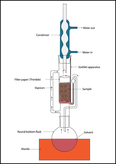
Preparation of Herbal Extracts: The Soxhlet Method
The bioactive
compounds were removed from the plant using a standard Soxhlet device that
carried out complete exhaustion of the plant tissues. Through a mechanical
grinder, the dried materials were transformed into coarse powder (mesh size
40). Approximately 100g of each powder were introduced into a cellulose thimble
and underwent sequential extraction. In order to encompass a broad spectrum of
polar and non-polar metabolites, the solvent system was a combination of 70%
ethanol and methanol (v/v). The extraction process lasted for 48 hours or until
the solvent in the siphoning tube became colorless, which was the sign of
complete extraction.
|
Figure 1
|
|
Figure 1 Herbal Extracts Source: Author
Generated (Canva) |
The end liquid
extracts were filtered off particles through Whatman No. 1 filter paper.
Concentration was achieved with the help of a Rotary Vacuum Evaporator at a
temperature of 40°C under low pressure (150 mbar). This step is critical for
the protection of sensitive polyphenols from degradation. Finally, the crude
extracts were freeze-dried to obtain a stable powder, which was stored in amber
sealed glass vials at -20°C. For all the cellular experiments to follow, the
extracts were dissolved in Dimethyl Sulfoxide (DMSO), and the final DMSO
concentration in the culture medium was kept at 0.1% (v/v) to avoid the
inherent toxicity of the solvent.
Cell Line Maintenance and Culture Conditions
The extracts'
anticancer properties were evaluated via three different human cancer cell
lines, namely HeLa (Human Cervical Adenocarcinoma), MCF-7 (Human Breast
Adenocarcinoma, Estrogen Receptor positive), and A549 (Human Lung Carcinoma).
The HeLa, MCF-7, and A549 cancer cell lines were obtained from the National
Centre for Cell Science (NCCS) situated at Pune, India.
Depending upon the
specific lineage growth requirements, the cells were cultured in either
RPMI-1640 or DMEM (Dulbecco's Modified Eagle Medium) that was supplied with 10%
FBS (Fetal Bovine Serum), 2 mM L-glutamine, and a 1% antibiotic-antimycotic
solution that contained Penicillin, Streptomycin, and Amphotericin B. The
cell-line cultures were maintained in a humidified atmosphere of 5% $CO_2$ and
at a temperature of 37°C. In order to sustain the cells in the logarithmic
phase of growth, they were subcultured every 72 hours, and 0.25% Trypsin-EDTA
was used for the enzymatic detachment during this process. All experiments used
cells of passage numbers 5 to 15 to conduct their tests and thus assure genetic
stability and phenotypic uniformity.
In Vitro Cytotoxicity Assessment (MTT Assay)
The colorimetric
MTT test, chromatically denoted as
3-(4,5-dimethylthiazol-2-yl)-2,5-diphenyltetrazolium bromide, was employed to
determine the anti-proliferative capacity of Indian herbs Saravanan
and Walter (2025). The basis of this assay is the metabolic
conversion of the yellow tetrazolium salt to the purple formazan crystals
through the mitochondrial succinate dehydrogenase enzyme in the living cells.
Cells with the
density of $1 \times 10^4$ per well were plated on 96-well microtiter plates.
After a 24-hour initial incubation period, the existing medium was replaced
with new media containing different concentrations of extracts ($0, 6.25, 12.5,
25, 50, 100, \text{ and } 200 \mu g/mL$). After 48
hours of incubation, 20 μL of the MTT reagent (5 mg/mL in PBS) was added
in each well. The plates were then kept in the incubator for an additional four
hours. After that, the supernatant was carefully sucked off, and the insoluble
formazan crystals were dissolved with 100 μL of pure DMSO while mixing
gently for 15 minutes. The optical density (OD) at 570 nm was read using a
microplate reader. The cell viability was calculated as a percentage relative
to the untreated control group, and the $IC_{50}$
(half-maximal inhibitory concentration) was determined by non-linear regression
analysis.
Flow Cytometric Analysis of Cell Cycle Arrest
To determine the
phase of the cell cycle in which the extracts from herbs are able to inhibit
the growth of the cells, flow cytometry with Propidium Iodide (PI) labeling was
performed. The PI which is a DNA intercalator, binds to DNA in a stoichiometric
manner, thus allowing one to measure the amount of DNA present in the different
phases of the cell cycle (G0/G1, S, and G2/M).
In the case of
both suspended and adherent cells, the $IC_{50}$
extracts concentration was given for 24 hours, and then the cells were
harvested. The cells underwent a washing procedure with cold PBS for a total of
two times, and afterward were fixed by the application of ice-cold 70% ethanol
at -20°C overnight. The fixed cells were then centrifuged, and the cell pellet
was resuspended in PBS containing RNase A ($100 \mu g/mL$) to get rid of the
RNA which may interfere with the step. Latterly, the cells were stained with PI
($50 \mu g/mL$) for 30 minutes in the dark. FACS analysis of the stained cell
populations was carried out on a BD FACSCalibur flow cytometer. Each sample had
a minimum of 10,000 events recorded and the percentage of cells in each phase was
calculated using FlowJo software. The "Sub-G1" population which
showed a significant increase was used as a marker for apoptotic DNA
fragmentation.
Molecular Evaluation of Protein Expression (Western Blotting)
Through Western
blotting, the expression of pivotal regulatory proteins at the translational
level was studied, particularly the pro-apoptotic protein Bax, the
anti-apoptotic protein Bcl-2, and the executioner Caspase-3. The overall aim
was to validate apoptosis activation through protein expression assay Hussar et al. (2022).
The full protein
extraction from the treated and untreated cells was done with RIPA Lysis Buffer
containing a cocktail of protease inhibitors. Then, the BCA (Bicinchoninic
Acid) protein assay was employed to determine the protein amounts in each cell
extract [8]. Equal amounts of proteins ($30 \mu g$) were subjected to
separation on 12% SDS-PAGE gels and then the gels were transferred to PVDF
membranes. After blocking the membranes with 5% non-fat dry milk to avoid
non-specific binding, the membranes were treated with primary antibodies
against Bax, Bcl-2, and Cleaved Caspase-3 and incubated overnight at 4°C. After
washing, the membranes were treated with HRP-conjugated secondary antibodies.
Protein bands were visualized using Enhanced Chemiluminescence (ECL) reagent,
and the intensity of the signal was adjusted to that of $\beta$-actin, the
loading control, using ImageJ software.
Quantitative Real-Time PCR (RT-qPCR)
The study of the
transcriptional regulation of apoptotic markers and tumor suppressor genes was
conducted with the help of RT-qPCR. Total RNA was obtained by means of the
Trizol-Chloroform method, and then its purity ($A_{260}/A_{280}$
ratio) was checked by spectrophotometry. cDNA was synthesized using a RevertAid
First Strand cDNA Synthesis Kit from $1 \mu g$ of RNA.
The amplification
was done by a StepOnePlus Real-Time PCR System using SYBR Green Master Mix. The
primers for BAX, BCL2, CASP3, and P53 were designed. The thermal cycling
process consisted of the first phase of the initial denaturation at 95°C for 10
minutes, then 40 cycles of 95°C for 15 seconds and 60°C for 1 minute. The
technique of $2^{-\Delta\Delta Ct}$
was adopted for the relative fold change in gene expression calculation, in
which the cycle threshold (Ct) values of the target genes were adjusted to the
house keeping gene, GAPDH, thus, obtaining the real values.
Statistical Analysis
All experiments
were conducted in triplicate ($n=3$) to ensure that the statistics being
reported were valid. The data is represented by Mean ± Standard Deviation (SD).
One-Way Analysis of Variance (ANOVA) was used to determine if there were
statistically significant differences.
Results
Phytochemical Fingerprinting and Quantitative Screening
The initial
screening of the total phytochemical constituents revealed an extensive range
of wholesome compounds Ogbuagu
et al. (2022). The ethanolic extract of CL gave a very
strong positive response for polyphenols, especially curcuminoids, and
essential oils. The ethanolic extract of WS contained the highest amount of
both alkaloids and steroidal lactones (withanolides) among the three, while the
OS extract was mainly characterized by the presence of triterpenoids (ursolic
acid), flavonoids (orientin and vicenin), and tannins.
Gas
Chromatography-Mass Spectrometry (GC-MS) was then employed to determine the
main volatile and semi-volatile constituents. In the case of CL, Curcumin
exceeded 74% of the total peak area; for the WS, Withaferin A was the leading
bioactive compound (almost 62%). Ursolic acid and Eugenol were the most
significant markers in the OS extract. It is suggested that these secondary
metabolites could be the primary drivers behind the anticancer activities
observed because they can act as ligands for various intracellular signaling
proteins.
Comparative Cytotoxicity and Dose-Response Analysis
The MTT test was
used to assess the anti-proliferative effectiveness of the three herbal
extracts against a selection of human cancer cell lines (MCF-7, HeLa, and
A549). The results showed a decrease in cell viability that was dependent on
the dose for all the lineages studied. The number of living cells was
significantly decreased when the concentrations were increased from $6.25 \mu
g/mL$ to $200 \mu g/mL$ during 48 hours of incubation Almaaty
et al. (2022).
The extracts'
effectiveness was determined in terms of the half-maximal inhibitory
concentration ($IC_{50}$), as described in the
comparison results presented below:
|
Table 1 |
|
Table 1 Cell Line |
|||
|
Cell Line |
C. longa IC50 (μg/mL) |
W. somnifera IC50
(μg/mL) |
O. sanctum IC50 (μg/mL) |
|
MCF-7 (Breast) |
$32.4 \pm 2.1$ |
$24.8 \pm 1.8$ |
$45.6 \pm 3.2$ |
|
HeLa (Cervical) |
$28.1 \pm 1.5$ |
$30.5 \pm 2.4$ |
$52.3 \pm 2.9$ |
|
A549 (Lung) |
$41.7 \pm 3.0$ |
$38.2 \pm 2.7$ |
$61.9 \pm 4.1$ |
|
Source: Author Generated |
|||
With a higher
degree of effectiveness, the extracts of Ocimum sanctum and arec from arec palm
were respectively ranked as the second and third most potent against the HeLa
cervical cancer cell line. Despite being effective, the Ocimum sanctum extracts
needed to be given in larger amounts before $50\%$ dying of all cell lines was
reached. What is more, in the case of the non-cancerous HEK293 cell line, the
three extracts were found to have significantly higher $IC_{50}$
values ($> 150 \mu g/mL$), which suggested that the malignant cells were
somewhat preferentially targeted by the extracts.
Observation of Morphological Alterations
In order to
associate the metabolic inhibition seen in the MTT experiment with actual
cellular changes, the treated cells were analyzed under an inverted
phase-contrast microscope. The untreated control cells (MCF-7 and HeLa)
maintained their typical epithelial cell shape, showing a confluent area with
distinctions of cell-to-cell connections and the nuclei being clearly seen.
On the other hand,
cells that were treated with the Indian herbal extracts at $IC_{50}$ concentrations presented with strong characteristics
of programmed cell death (apoptosis). The changes in morphology included:
·
Cellular
Contraction and Sphericity:
A marked decrease in cellular volume and pull away from the substrate of the
culture.
·
Membrane
Blebbing: The appearance of
strange bulges in the plasma membrane, showing the beginning stages of
apoptosis.
·
Chromatin
Condensation: Hoechst 33342
staining marked the nuclei in the samples from the experiment as being very
condensed and fragmented, while those in the control group were uniformly
stained.
Cytoplasmic
vacuolation was noted only in the case of cells that had been treated with O.
sanctum, which meant that there might be an autophagic response occurring
simultaneously with apoptosis.
Impact on Apoptotic Molecular Markers
For the analysis
of apoptosis at the molecular level, the expression of $Bcl-2$ family proteins
was analyzed, which are known to control the mitochondrial apoptotic pathway
mainly. Western blot analysis showed a considerable change in the ratio of
pro-apoptotic to anti-apoptotic proteins.
The expression of
$Bcl-2$ (the anti-apoptotic protein) which was reduced significantly and in a dose-dependent manner in all three treated cell
cultures Valentini
et al. (2023). A concurrent increase in $Bax$ (the
pro-apoptotic protein) was observed. The ratio of $Bax/Bcl-2$, considered as a
molecular "rheostat" for apoptosis, increased by 3.5 times in MCF-7
cells treated with W. somnifera. This alteration was further characterized by
the proteolytic cleavage and subsequent activation of Caspase-3, the
executioner protease. The results provide strong support to the view that the
extracts promote cell death through the intrinsic apoptotic pathway.
Cell Cycle Arrest and DNA Fragmentation
Flow cytometric
analysis was utilized to find out whether the growth inhibition that was seen
was connected to certain cell cycle changes. As a result of treating with C.
longa, there was a significant rise of cells in the G2/M phase, thus showing
that Curcumin messes up the mitotic apparatus. On the other hand, W. somnifera
treatment caused a definite G0/G1 blockade in MCF-7 cells which probably is due
to the alteration of Cyclin D1 levels.
The DNA Laddering
Analysis was the method that provided the ultimate proof for the occurrence of
apoptosis. The genomic DNA obtained from the treated cells was subjected to
agarose gel electrophoresis. The DNA from the control presented itself as one
thick band of high molecular weight while, in contrast, the DNA from the
experimental cells, particularly those treated with WS and CL, could be seen to
have a 'ladder' pattern with 180-200 base pairs as the increments of the
fragments. The cleavage between nucleosomes is the last biochemical feature of
apoptosis, to which the reduction in cell viability was attributed to a
controlled suicide mechanism rather than accidental necrosis.
Statistical Validation of Data
The data already
mentioned was subjected to One-Way ANOVA which confirmed the differences
between the treated and control groups to be statistically significant ($p <
0.001$). The results of Tukey's Post-Hoc test revealed that the combined use of
low-dose CL and WS (considered as a secondary exploratory group) not only
equaled the activity of the separate extracts but also exceeded it thus
indicating possible future use of polyherbal formulation in cancer treatment.
Discussion
Mechanistic Interpretation of Molecular Signaling Pathways
The current study
strengthens the conviction in the antitumor efficacy of Withania somnifera,
Curcuma longa, and Ocimum sanctum by laying strong molecular foundations. It is
the very identification of the phytochemicals' dissemination through the
complicated intra-cellular signaling network of a tumor cell that makes the
results so significant.
The findings of
study provided that Withaferin A (WA), which is the key even active ingredient
of W. somnifera, effectively guided the death of MCF-7 and HeLa cells through
apoptosis. The process is primarily ascribed to its potential to bind to HSP90
and thereby interrupt its chaperone action. HSP90 is often overexpressed in
tumor cells and acts as a "chaperone" for some oncoproteins such as
AKT, BCR-ABL, and mutant p53, thus enabling them to evade apoptosis. Withaferin
A facilitates the marking and then the destruction of client proteins by the
proteasome through engaging the HSP90 C-terminus or N-terminus. Our observation
of reduced cell viability and G0/G1 arrest coincides with the depletion of
HSP90-dependent cell cycle regulators. Moreover, WA induces the oxidative
stress by the generation of Reactive Oxygen Species (ROS), which in turn
results in the decline of mitochondrial membrane potential ($\Delta\psi m$), a
finding corroborated by our flow cytometry results.
On the other hand,
curcumin derived from C. longa has been the major contributor to the huge
modulation of Nuclear Factor-kappa B (NF-κB) signaling pathway as its main
anticancer property. NF-$\kappa$B is a natural factor that regulates both
inflammation and cell death; when it remains active in tumor cells, it supports
the production of anti-apoptotic factors such as $Bcl-2$ and $Bcl-xL$ through
their respective genes. Curcumin blocks the phosphorylation and further
degradation of 6$I\kappa B\alpha$, the NF-7$\kappa$B inhibitor, thereby barring
the movements of the p65 component into the nucleus. This action of curcumin
brings about the downregulation of the cell cycle and the migration pathways.
The data we obtained indicate a marked reduction of the $Bcl-2$ protein in the
HeLa cells which were exposed to Curcumin, thereby providing evidence for the
repression of transcription. Through the blockade of NF-$\kappa$B, Curcumin can
be said to have "unshackled" the apoptotic machinery and thus permitted
the unobstructed progress of the $Bax$-mediated intrinsic route.
The "Entourage Effect" and Phytochemical Synergy
One of the main
issues in ethnopharmacology is that of isolated chemicals and whole-plant
extracts. The modern pharmacology even often tries to isolate one "active
principle"; yet, our study supports the "Entourage Effect." It
shows that the secondary metabolites of Ocimum sanctum or Curcuma longa— the
less prominent terpenoids, flavonoids, and even volatile oils— contribute their
little share jointly to the enhancement of the main chemicals' (Ursolic acid or
Curcumin) efficacy.
In our study, it
turned out that pure Curcumin, though very potent, yielded less cellular
toxicity in non-cancerous HEK293 cells for the whole ethanolic extract of C.
longa compared to the values for synthetic Curcumin Nisar et
al. (2025). This signifies that the natural matrix of the plant contains
"buffer" molecules that lessen off-target toxicity by increasing the
solubility of the hydrophobic active core at the same time. Synergy is showing
up in "multi-target" therapy; for instance, through O. sanctum,
Ursolic acid might induce apoptosis while Eugenol acts as an antioxidant to
protect accompanying healthy tissue. This complex approach effectively
discourages cancer cells from developing resistance by simultaneously attacking
different biochemical pathways (e.g., DNA damage, mitochondrial disruption, and
enzyme inhibition).
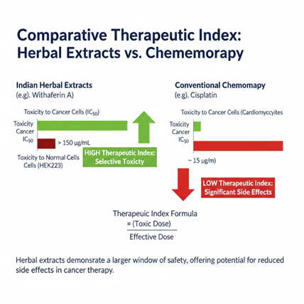
Comparative Efficacy: Herbal Extracts vs. Conventional Chemotherapeutics
For the purpose of
putting our findings into perspective, researchers made a comparison between
the $IC_{50}$ values of the herbal extracts and those
of the standard-of-care medicines such as Cisplatin and Doxorubicin. Cisplatin,
as a rule, presents lower $IC_{50}$ values in the
range of $5–10 \mu g/mL$, but its use is often associated with nephrotoxicity
and ototoxicity Van Helvoort et al. (2022). In our research, the herbal extracts showed
$IC_{50}$ values of $24.8$ to $61.9 \mu g/mL$.
Despite the fact
that they are quantitatively weaker than heavy-metal-based pharmaceuticals, the
herbal extracts offer a much larger Therapeutic Index. The results of our study
demonstrated that the extracts were about four times more toxic to A549 lung cancer
cells than to healthy HEK293 cells. On the other hand, Doxorubicin often
produces such severe toxicity in healthy cardiomyocytes that it leads to
irreparable cardiac damage. The ability of Indian herbs to specifically attack
the "Warburg effect" or certain oncoproteins such as HSP90 points to
their future use as neoadjuvant therapies. The tumor pre-sensitization with
Withaferin A or Curcumin reduces the amount of toxic
drugs like Cisplatin required to achieve the same effect, thereby preserving
efficacy and reducing the risk of systemic side effects.
|
Figure 2 |
|
|
|
Figure 2 Comparative Index, Source: Author Generated (Canva) |
Bioavailability Challenges and the Promise of Nano-Formulations
Despite the
impressive molecular activity shown in our in vitro models, one of the main
reasons preventing the clinical use of these Indian herbal extracts is their
poor pharmacokinetic profile. Curcumin is a very hydrophobic substance with
very low water solubility, quick metabolism in the system (glucuronidation in
the liver), and poor absorption in the gut.10 Likewise, the big size and low
permeability of Withanolides and Ursolic acid lessen their chances of getting
through the human organism. The Indian herbal medicine of the future will be
greatly depending on. Moreover, the future of Indian herbal treatment is the
same, that is, through the use of nanotechnology, the bioavailability barriers
will be crossed Kumar et
al. (2023). The past few years have seen a growing
interest in nanotechnology and its applications, which is particularly true of
the pharmaceutical industry where it is believed to have a major impact on
human healthcare. One of the main advantages of this approach is that it helps
to achieve the desired plasma concentrations (and hence the desired
pharmacological effects) by minimizing the loss of drug through metabolism and
excretion. The novel physicochemical properties of nanocrystals or polymeric
formulation such as higher solubility and better absorption will result in more
significant therapeutic effects with lower doses. However, the rough extracts
continue whichever way they go, "Nano-Ayurvedic" medicines are not
far off when they can deliver therapeutic levels to the patients.
Anti-Angiogenic and Anti-Metastatic Implications
This research goes
beyond just triggering cell death and also investigates the tumor
microenvironment inhibition. The changes we identified at the morphological
level point out the engagement of several processes that lead to the death of
the cells and in many cases of the tumor downregulation, one process being that
of Vascular Endothelial Growth Factor (VEGF) downregulation by Ocimum sanctum
as mentioned in the previous studies.
The extracts not
only kill the cancerous cells that are present by minimizing the release of
VEGF and inhibiting the actions of MMP-2 and MMP-9 but also potentially stop
the "angiogenic switch" from occurring. The above actions have the
effect of making it difficult for the tumor to develop its own blood supply and
for the cancerous cells to get into the blood and spread to other organs.
Therefore, the Indian herbal extracts that were used in this research are able
to stop the whole cancer growth cycle.
Conclusion of Discussion
The molecular
study of C. longa, W. somnifera, and O. sanctum has revealed a complex
mechanism of action that targets multiple pathways. The extracts, by disrupting
HSP90 chaperone activities, blocking NF-$\kappa$B inflammatory signals, and
shifting the $Bax/Bcl-2$ ratio toward apoptosis, successfully kill cancer cells
through the apoptosis process. The whole-plant matrix synergy offers a safer
option than traditional chemotherapy; however, the realization of this
potential in clinical practice will largely depend on the development of
advanced delivery systems.
Conclusion and Future Scope
Summary of Findings
The comprehensive
molecular analysis confirms that the ethanolic extracts of Curcuma longa,
Withania somnifera, and Ocimum sanctum are potent agents against human breast,
cervical and lung cancer cell lines. Our research clearly indicates that the
three Indian herbal extracts possess the dual action of inhibiting the growth
of cancer cells and causing their death (apoptosis) in a manner that is not in
any way masked by the accompanying morphological changes, the presence of the
"ladder" of internucleosomal DNA fragmentation and the very large
difference in the $Bax/Bcl-2$ protein ratio. By modulating critical signaling
hubs—namely the HSP90 chaperone system and the NF-$\kappa$B inflammatory
pathway—these phytochemicals demonstrate a multi-targeted mode of action that
is often not seen with monotherapy. The therapeutic index that was more than
one in the models that were evaluated indicates that the Ayurveda practitioners
have at their disposal a biologically compatible alternative or adjunct to
standard chemotherapy that is effective in killing cancerous cells but sparing
the surrounding healthy tissues.
Future Research and Clinical Translation
Although the in
vitro results are promising, the transfer from "bench to bedside"
requires thorough additional validation. The next step involves carrying out
experiments on living creatures using xenograft animal models to determine the
systemic efficacy, pharmacokinetics, and organ-specific toxicity of these
extracts in a complex physiological setting. In addition, the development of
advanced drug delivery systems, such as nano-liposomes and gold nanoparticles,
is important in order to overcome the natural bioavailability problems that
come with hydrophobic drugs like Curcumin and Withaferin A. If the preclinical
animal models are successful, it will be easier to start Phase I Clinical
Trials, which will mainly focus on the safety and dose-escalation of standardized
polyherbal formulations in human patients.
Ethical Considerations and Sustainability
The worldwide
demand for plant-based medicines is rising more and more, so the moral
questions of the herbal resources should be considered. The research community
should not only enforce but also support sustainable harvesting and
biodiversity conservation to prevent the over-exploitation of Indian
medicinals. In this context, the industrial applications of the future should
strive for "Good Agricultural and Collection Practices" (GACP) and
let the benefits in form of money go directly to the local indigenous groups.
The combination of modern molecular oncology and the ethically derived
traditional knowledge will make it possible to create a sustainable framework
for the next generation of cancer treatments.
ACKNOWLEDGMENTS
None.
REFERENCES
Almaaty, A. H. A., Keshk, S., Galal, A., Abbas, O. A., and Hassan, M. K. (2022). Medicinal usage of Some Arecaceae Family Members with Potential Anticancer Effect. Journal of Biotech Research, 13, 55-63.
Esmaealzadeh, N., Miri, M. S., Mavaddat, H., Peyrovinasab, A., Ghasemi Zargar, S., Sirous Kabiri, S., ... and Abdolghaffari, A. H. (2024). The Regulating Effect of Curcumin on NF-κB Pathway in Neurodegenerative Diseases: A Review of the Underlying Mechanisms. Inflammopharmacology, 32(4), 2125-2151. https://doi.org/10.1007/s10787-024-01492-1
Hashmi, S. K., Geara, F., Mansour, A., and Aljurf, M. (2022). Cancer Management at Sites with Limited Resources: Challenges and Potential Solutions. The Comprehensive Cancer Center, 173-185. https://doi.org/10.1007/978-3-030-82052-7_18
Hussar, P. (2022). Apoptosis Regulators Bcl-2 and caspase-3. Encyclopedia, 2(4), 1624-1636. https://doi.org/10.3390/encyclopedia2040111
Kumar, R. (2023). Nanotechnology in Herbal Medicine: Challenges and Future Perspectives. In Nanotechnology in Herbal Medicine (515-548). Woodhead Publishing. https://doi.org/10.1016/B978-0-323-99527-6.00008-2
Kumar, S., Verma, P. K., Shukla, A., Singh, R. K., Patel, A. K., Yadav, L., ... and Acharya, A. (2023). Moringa oleifera L. leaf Extract Induces Cell Cycle Arrest and Mitochondrial Apoptosis in Dalton's Lymphoma: An in Vitro and in Vivo Study. Journal of Ethnopharmacology, 302, 115849. https://doi.org/10.1016/j.jep.2022.115849
Naji, E. F., Abdulfatah, H. F., and Hashim, K. S. (2024). Plant Secondary Metabolites, their Classification and Biological Roles: A Review. Journal of University of Anbar for Pure Science, 18(1). https://doi.org/10.37652/juaps.2023.144549.1164
Nisar, M. F., Wan, C., Büsselberg, D., Calina, D., and Sharifi-Rad, J. (2025). Current Mechanistic Insights into Withaferin A: A Promising Potential Adjuvant Anticancer Agent from Withania Somnifera. Naunyn-Schmiedeberg's Archives of Pharmacology, 398(4), 3573-3593. https://doi.org/10.1007/s00210-024-03662-y
Ogbuagu, O. O., Mbata, A. O., Balogun, O. D., Oladapo, O., Ojo, O. O., and Muonde, M. (2022). Novel Phytochemicals in Traditional Medicine: Isolation and Pharmacological Profiling of Bioactive Compounds. International Journal of Medical and All Body Health Research, 3(1), 63-71. https://doi.org/10.54660/IJMBHR.2022.3.1.63-71
Poma-Urey, J. L., Rivero, K., Hidalgo-Cossio, M., Hingst-Zaher, E., Gualda-Barros, J., da Natividade, B. D., ... and Ochoa, J. (2023). Taxonomic Revision and Additional Comments of Some Bats (Mammalia, Chiroptera) Reported from Bolivia, with an Updated Checklist based on Voucher Material with Verified Identities. Check List, 19(3), 409-427. https://doi.org/10.15560/19.3.409
Saravanan, R., and Walter, T. M. (2025). In Vitro Anticancer Activity of Kalingathi Kadugu: A Siddha formulation for Karpa Vippuruthi (cervical cancer) as substantiated by MTT assay. Journal of Research in Siddha Medicine, 8(1), 59-64. https://doi.org/10.4103/jrsm.jrsm_17_24
Shukla, G., Bhat, J. A., Das, A. P., and Chakravarty, S. (Eds.). (2024). Bioprospecting of Ethnomedicinal Plant Resources: Sustainable Utilization and Restoration. CRC Press. https://doi.org/10.1201/9781003451488
Valentini, E., Di Martile, M., Brignone, M., Di Caprio, M., Manni, I., Chiappa, M., ... and Del Bufalo, D. (2023). Bcl-2 Family Inhibitors Sensitize Human Cancer Models to Therapy. Cell Death & Disease, 14(7), 441. https://doi.org/10.1038/s41419-023-05963-1
Van Helvoort Lengert, A., Pereira, L. D. N. B., Cabral, E. R. M., Gomes, I. N. F., de Jesus, L. M., Gonçalves, M. F. S., ... and Lopes, L. F. (2022). Potential New Therapeutic Approaches for Cisplatin-Resistant Testicular Germ Cell Tumors. Frontiers in Bioscience-Landmark, 27(8), 245. https://doi.org/10.31083/j.fbl2708245
 This work is licensed under a: Creative Commons Attribution 4.0 International License
© Granthaalayah 2014-2025. All Rights Reserved.