Introducing Optical Microscopy Methodology Displaying Emitted Electromagnetic Energy of Pre-cancerous Skin Squamous Cell Carcinoma
Abrahám A. Embí 1![]()
![]()
1 BS MBA,13442 SW 102 Lane Miami, 33186, Florida, United States
|
|
|
ABSTRACT |
|
|
Ever since the development in 2015 (Embi et al. (2015), Scherlag et al. (2015)) and published in 2016 Scherlag et al. (2016) of a tabletop microscopy methodology aimed to record plant and animal tissue energy in the form of electromagnetic radiation (EMR), this author and others have had the opportunity to explore and publish in the less traveled road of Biophysics. This was mainly done by using an easily accessible human miniorgan, namely a hair follicle Schneider et al. (2009). |
|||
|
Received 20 December 2022 Accepted 22 January 2023 Published 06 February 2023 Corresponding Author Abrahám A. Embí, Embi21@att.net DOI 10.29121/granthaalayah.v11.i1.2023.4949 Funding: This research
received no specific grant from any funding agency in the public, commercial,
or not-for-profit sectors. Copyright: © 2023 The
Author(s). This work is licensed under a Creative Commons
Attribution 4.0 International License. With the
license CC-BY, authors retain the copyright, allowing anyone to download,
reuse, re-print, modify, distribute, and/or copy their contribution. The work
must be properly attributed to its author.
|
|||
|
Keywords: Cancer Tissue Energy, Potassium
Ferricyanide, Anisotropy, Precancerous Tissue, Squamous Cell Biopsy,
Bioelectromagnetic Energy DEFINITIONS OF TERMS Anisotropy: “(of an object or substance) having a physical property that has a different value when measured in different directions. A simple example is wood, which is stronger along the grain than across it”. (Oxford Dictionary). EMR: Electromagnetic Radiation. K3Fe: Acronym for Potassium Ferricyanide K₃ [Fe (CN)₆] crystals. Scab: Protective tissue covering those forms after your skin has been damaged. SSP: Single Slide Preparation. Sample placed in center of glass slide and covered by two drops K3Fe. Allowed to evaporate.
|
|||
1. INTRODUCTION
Ever since the development in 2015 (Embi et al. (2015), Scherlag et al. (2015)) and published in 2016 Scherlag et al. (2016) of a tabletop microscopy methodology aimed to record plant and animal tissue energy in the form of electromagnetic radiation (EMR), this author and others have had the opportunity to explore and publish in the less traveled road of Biophysics. This was mainly done by using an easily accessible human miniorgan, namely a hair follicle Schneider et al. (2009).
2. AREAS EXPLORED
Using the tabletop microscopy was instrumental in proposing endogenous EMRs emitted during cell respiration as an additional factor in cancer genesis Embi (2016). The latter was supported by prior in vitro findings where the breakdown of the H2O2 during cellular respiration oxidation reduction reaction was demonstrated to penetrate a 1 mm glass slide causing disruption of crystallized Prussian Blue Stain crystals in solution. The latter finding was then rationalized as an EMR foundation for the hypothesis Embi (2016). Other publications introduced this author to the concept of anisotropy, being a non-uniform wave of energy activation Moscow Institute of Physics and Technology. (2016). The importance of anisotropy is identified as an essential supporting mechanism in this research letter, Why? Because the anisotropic Potassium Ferricyanide of formula K₃[Fe(CN)₆] in solution has been established as partially absorbing incoming EMRs (8,9) and the 2016 published optical microscopy methodology includes K₃[Fe(CN)₆] in solution plus very small iron particles; for lack of resources this homebound citizen scientist has been using diluted K₃[Fe(CN)₆] in solution as sole component with excellent results recording the tissue absorption of emitted EMR.
3. COMPARATIVE RESULTS RATIONALE
The results and images herein presented are comparative, meaning experiments done on normal and precancerous tissues are mounted and analyzed using the same water drops diluted Potassium Ferricyanide crystals. As a clarifying statement: For the purpose of this presentation the fact that at present K₃[Fe (CN)₆] has been proven to partially absorb incoming EMRs is a moot point, Why? Because both precancerous and normal tissue samples were analyzed using crystals from the same badge (Exhibit 1, Exhibit 2).
4. Prior Publication Comparing Normal and Squamous Cell Carcinoma Scabs
As fate has it, this author had compared and published images of EMR emissions of normal vs post-biopsied positive squamous cell carcinoma scabs. In that paper, images presented showed a notable difference in emissions between the normal and cancerous scabs Embi (2022). Not satisfied with the experiment again, upon a scheduled dermatologist appointment to freeze some pathologists interpreted pre-cancerous lesions, I asked if I could get tissue samples before freezing, the pre-cancerous lesion was shaved, and the tissue samples placed in a small container for further analysis. The normal epidermal tissue samples were retrieved via tweezers from this 80 y/o skin.
EXHIBIT I
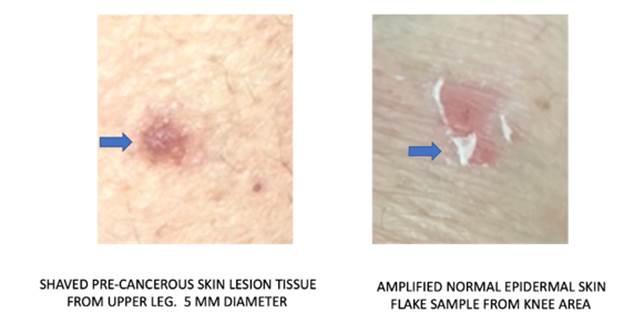
|
Exhibit 1 Showing Tissue Selection |
5. The Results
A pinch or Potassium Ferricyanide crystals placed on a glass slide and diluted in three drops of bottled water. The normal tissue and the cancerous tissue small fragments were placed at opposite ends of a glass slide containing the testing solution and allowed to evaporate. After and during evaporation images were recorded in a video microscope and store in an Apple Computer video application for later retrieval. A total of three pre-cancerous experiments were done. Important to note that the tissue donor (myself) have had several Squamous Cell Skin Carcinoma removed.
EXHIBIT II
|
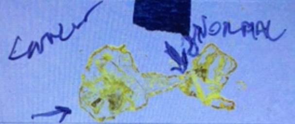
Exhibit 2 Slide Image Post
Evaporation Demonstrating Using Same Potassium Ferricyanide Solution for Both
Samples |
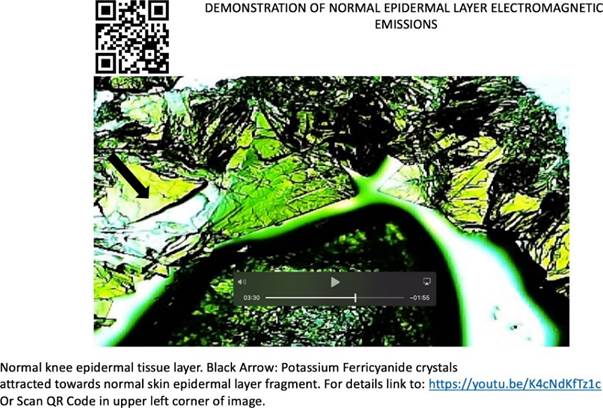
The image below (Exhibit 3) is representative of the slower interaction (absorption of EMRs) between Potassium Ferricyanide in solution and a normal epidermal skin flake (See Exhibit 1 above). Pleas compare the video images in both exhibits to appreciate the observed exacerbated EMR emissions in a pre-cancerous lesion.
EXHIBIT III
|
Exhibit 3 Demonstration of Normal Epidermal Layer Electromagnetic Emissions |
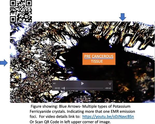
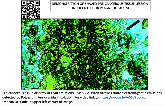
The image and video below (Exhibit 4) were n=1 of the precancerous tissue biopsy placed on a slide and covered by drops of Potassium Ferricyanide in solution. Please notice the increased speed in crystals formation, indicative of a strong electromagnetic environment. This exacerbated speed was not present in the normal skin epidermal tissue. This increased speed was also not appreciated in two of the pre-cancerous samples n=2 and n=3. Possibly corroborating the presence of multiple EMR foci in pre- cancerous tissue. (Compare videos presented in Exhibits)
EXHIBIT IV

|
Exhibit 4 Demonstration of Shaved Pre-Cancerous Tissue Lession Induced Electromagnetic Storm |
EXHIBIT V
|
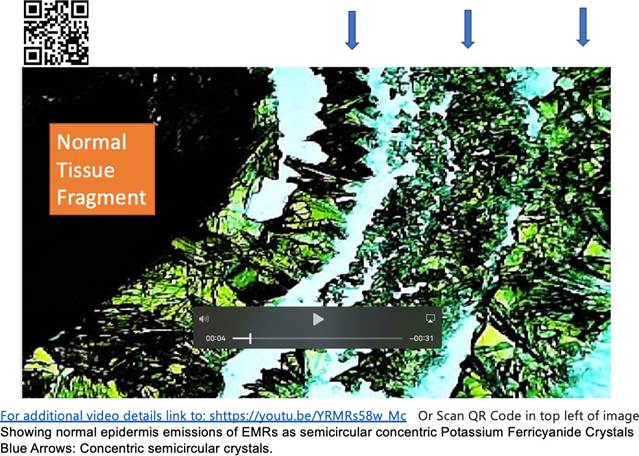
Exhibit 5 Example of Normal Tissue Emitted Energy |
EXHIBIT VI
|
Exhibit 6 Example of Pre-Cancerous Tissue Chaotic Energy Emission |
EXHIBIT VII
|
Exhibit 7 Another Example of Pre-Cancerous Tissue Electromagnetic Emission |
6. SUMMARY
In closing, in vitro experiments from two different papers, in the present submission and in reference Embi (2022) published in your journal are presented confirming the presence of aberrant or fragmented EMR emissions in pre-cancerous tissue.
CONFLICT OF INTERESTS
None.
ACKNOWLEDGMENTS
None.
REFERENCES
Baranov, D. G., Edgar, J. H., Hoffman, T., Bassim, N., and Caldwell, J. D. (2015). Perfect Interferenceless Absorption at Infrared Frequencies by a Van Der Waals Crystal. Physical Review B, 92(20). https://doi.org/10.1103/PhysRevB.92.201405.
Embi, A. A. (2016). Cellular Respiration Oxidation Reduction Reactions Electromagnetic Fields Emissions as Possible Causative Agent in Diseases: A Chronic Bombardment Theory. Physics Journal, 2(3), 226–230.
Embi, A. A. (2016). Endogenous Electromagnetic Forces Emissions During Cell Respiration as Additional Factor in Cancer Origin. Cancer Cell International, 16, 60.
Embi, A. A. (2022). Detecting and Displaying Energy from Skin Cancer Lesions Comparison of Post Biopsy Skin Cancer Scabs with Normal Skin Injury Scabs. Biophysics approach. International Journal of Research -Granthaalayah, 10(6), 25–32.
Embi, A.A., Jacobson, J.I., Sahoo, K., and Scherlag, B. J. (2015). Demonstration of Inherent Electromagnetic Energy Emanating from Isolated Human Hairs. Journal of Nature and Science, 1(3): e55.
Figgis, B. N., Gerloch, M., Mason, R., and Sydney, R. (1969). Nyholm the Crystallography and Paramagnetic Anisotropy of Potassium Ferricyanide. https://doi.org/10.1098/rspa.1969.0031.
Moscow Institute of Physics and Technology. (2016). New Way to Absorb Electromagnetic Radiation Demonstrated: Scientists Show That it is Possible to Fully Absorb Electromagnetic Radiation Using an Anisotropic Crystal. Sciencedaily. 2022, December 13.
Scherlag, B. J., Huang, B., Zhang, l., Sahoo, K., Towner, R., Smith, N., Embi, A. A., and Po, S. S. (2015). Imaging the Electromagnetic Field of Plants (Vigna radiata) Using Iron Particles: Qualitative and Quantitative Correlates. Nature and Science, 1(3), e6.
Scherlag, B. J., Sahoo, K., Abraham, A., and Embi, A. (2016). Novel and Simplified Method for Imaging the Electromagnetic Energy in Plant and Animal Tissues. Nanoscience and Nanoengineering, 2(1), 6–9.
Schneider, M. R., Schmidt-Ullrich, R., and Paus, R. (2009). The Hair Follicle as a Dynamic Miniorgan. Current Biology, 19(3), R132–R142. https://doi.org/10.1016/j.cub.2008.12.005.
 This work is licensed under a: Creative Commons Attribution 4.0 International License
© Granthaalayah 2014-2023. All Rights Reserved.