Spontaneous Levitation
of Plucked Human Hairs on a Glass Slide when Immersed in Liquid Potassium
Ferricyanide.
Supporting a Tabletop Microscopy Methodology for the imaging of Electromagnetic Energy
in Plant and Animal tissue
Abraham A. Embi 1![]()
![]()
1 BS MBA,13442 SW 102 Lane Miami, 33186,
Florida, United States
|
|
|
ABSTRACT |
|
|
“in a seminal paper describing the origin of magnetic fields in the human body Cohen et al. (1980) by using sophisticated equipment stated: “Most of the field over the head is produced by electrical sources associated with the hair follicles of the scalp; this field is produced only as a response to touching or pressing the scalp in regions where the hair is dense”. Recently, an optical microscopy method was developed in 2015 and published a year later Scherlag et al. (2016) that also detects bioelectromagetic fields from hair follicles”. |
|||
|
Received 04 July 2022 Accepted 21 July 2022 Published 03 August 2022 Corresponding Author Abraham
A. Embi, embi21@att.net DOI 10.29121/granthaalayah.v10.i7.2022.4710 Funding: This research
received no specific grant from any funding agency in the public, commercial,
or not-for-profit sectors. Copyright: © 2022 The
Author(s). This work is licensed under a Creative Commons
Attribution 4.0 International License. With the
license CC-BY, authors retain the copyright, allowing anyone to download,
reuse, re-print, modify, distribute, and/or copy their contribution. The work
must be properly attributed to its author.
|
|||
|
Keywords: Potassium Ferricyanide, Magnetic Fields,
Electromagnetic Radiation In this manuscript additional information is
presented adding scientific support to the optical microscopy as described in
2016. |
|||
1. INTRODUCTION
The main purpose of this commentary is to introduce additional scientific evidence confirming previous findings whereby human hair follicles were documented emitting energy in the form of electromagnetic fields (EMFs). A methodology using Potassium Ferro or Ferricyanide was developed in 2015 and published in 2016 (36 years later). A simplified method accomplishing similar documentation of hair follicles EMFs was introduced approximately and labeled in part by the authors as a “novel tabletop optical microscopy methodology”.
2. POTASSIUM FERRICYANIDE AND TOTAL ABSORPTION OF INCOMING EMFS
For simplicity in this short report the acronyms K3Fe will be used instead of the conventional C6N6FeK3 formula. As fate has it, this citizen scientist became aware of an intrinsic property of K3Fe, which is that this cyano compound was found to “totally absorb incoming electromagnetic radiation” Figgis et al. (1969) Based on that information, this author could then fully understand and interpret the images obtained throughout previous years. An example of said image is shown below Figure 1 .
3. EXPECTED IMAGE OF A NON-LEVITATING HAIR IMMERSED IN LIQUID K3FE
Reproduced from: Abrahám A. Embí BS MBA. (2020). INTRODUCING CRYSTALLIZATION BACKWARD SUCTION TRAPPING LIPIDS AND DEBRIS AS PROPOSED ADDITIONAL FACTOR IN THE GENESIS OF CORONARY ARTERY DISEASE. International Journal of Research -GRANTHAALAYAH, 8(9), 215-233. https://doi.org/10.29121/granthaalayah.v8.i9.2020.1174
Figure 1
|
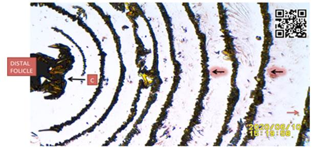
Figure 1 Human Scalp Hair SSP K3Fe Showing Crystallization Semicircles Reflecting Human Hair Bioelectromagnetic Waves Triggering Nucleation and Crystallization. C: Heavy Crystallization Caused by BSW Near the Follicle,
Highlighted Black Arrows: Pointing at K3Fe Crystals Delineating the Hair
Follicle Bio Electromagnetic Waves. Red Arrow: (Bottom right) Pointing at Hair
Follicle Magnetic Reach as Expressed by Crystallization. For Additional Details Link to: https://youtu.be/o1u5mHopdeo Or Scan QR Code in Right Upper Corner of Figure |
4. THE PROOF IS IN THE IMAGES
Over the years, also observed was an unusual pattern of K3Fe crystallization in some experiments. A different pattern of K3Fe crystals was detected; and only when the slide was handheld and viewed flat is that the lifting of mostly the distal hair follicle was identified as the culprit. In other words, when the hair tissue covered by K3Fe in solution remains in a flat position on the slide images as seen in Figure 1 above is the norm. When the hair was levitating a different crystallization pattern arose Figure 2 below.
5. TYPICAL PHOTOGRAPHIC IMAGE OF GLASS
SLIDE WITH LEVITATING DISTAL HAIR SEGMENT
Figure 2
|
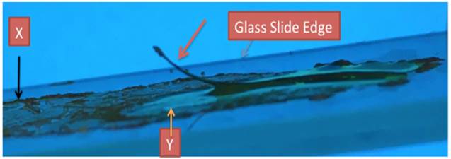
Figure 2 Lateral Photograph of
Slide Held in Front of Light from Video Monitor of Human Mustache Hair
Plucked and Immersed in Liquid K3Fe. X: Evaporated Potassium Ferricyanide Crystals Y: Area of Potassium Ferricyanide Not Exposed to
Hair Biomagnetism. Red Arrow: Untouched Lifted Hair. Please Compare Labels X,
Y With Figure 4 Below |
6. EXPERIMENT SHOWING LEVITATING HAIR IMMERSED IN LIQUID K3FE
Figure 3
|
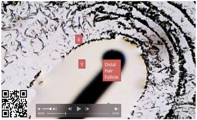
Figure 3 Frame 00:32 Showing Distal Human Hair on Liquid Solution of Potassium Ferricyanide (K3Fe). Hair Image Out of Focus Since It Is Not Directly in Contact with K3Fe. X: Potassium Ferricyande Crystals Detecting Hair Biomagnetism. Y: Liquid Potassium Ferricyanide. For Additional Details Please link to: https://youtu.be/7vZ_cGeGAzo…. Or Scan QR Code in image |
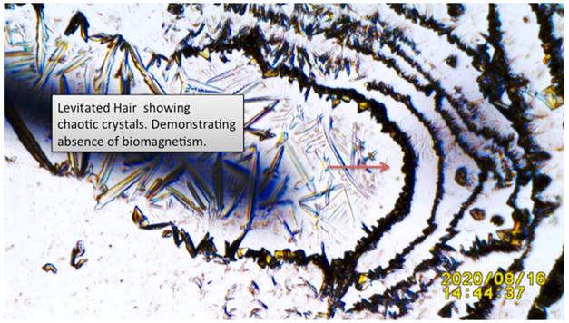
7. SAME EXPERIMENT AS Figure 3 (ABOVE) AFTER K3FE EVAPORATION
Figure 4
|
Figure 4 Showing Video Frame
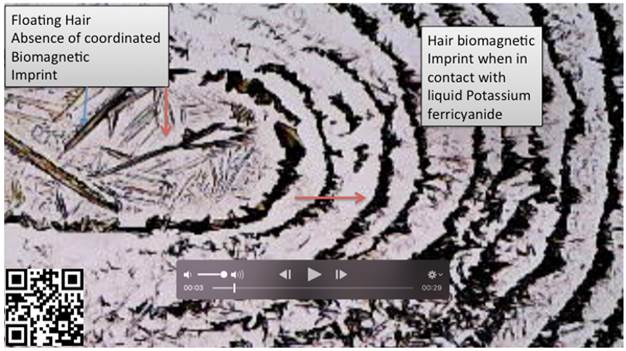
of Same Hair as Figure 3 Two Types of Crystals Shown: Flat Red Arrow:
When Hair in Touch with Solution Depicting Concentric Magnetic Waves Absorbed
by The Ferricyanide…. And: Vertical Red Arrow: Chaotic Irregular Ferricyanide
Crystals Theorized Caused by The Absence of a Biomagentic Source. for Details
Link To: https://youtu.be/7vZ_cGeGAzo …or link to QR Code in
left corner of image |
8. ANOTHER EXPERIMENT ALSO SHOWING LEVITATED HAIR ON SLIDE
Figure 5
|
Figure 5 Another Example from Another Hair Showing Different Crystallization Patterns When Hair Is Not in Touch with Slide (Levitating). Red Arrow: Showing Expected Concentric K3Fe Crystals Caused by Emfs from Hair |
9. ADDITIONAL EXPERIMENT DOCUMENTING ABSENCE OF METABOLISM UNABLE TO TRIGGER CRYSTALLIZATION. DEAD HAIR IN K3FE UNABLE TO TRIGGER K3FE CRYSTALS
Figure 6
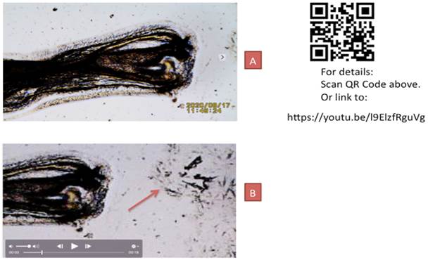
|
Figure 6 Additional Proof of Dead
Hair Follicle Unable to Emit Electromagnetic Radiation When Immersed in
Liquid K3Fe. A: Image of Dead Hair. B: Red Arrow- Same hair Immersed in K3Fe Unable
to Emit Concentric Potassium Ferricyanide Crystals |
10. BRIEF SUMMARY
Human Tissue Electromagnetism Confirmed by a “Novel Tabletop Microscopy Methodology”.
In this manuscript, the validity of a tabletop microscopy method to detect electromagnetic emissions from living tissue is reinforced. How? Since a picture is worth many words, images from In Vitro experiments by this author are presented. Again, the hair follicle was used as sentinel in documenting the accuracy of human tissue electromagnetic emissions shown via four provocative maneuvers. The first by our own hair tissue levitating in response to its immersion in liquid Potassium Ferricyanide (K3fe). The hair is shown photographically spontaneously levitating Figure 2 The same phenomena is shown under the microscope Figure 3 Figure 4 Figure 5 plus video) causing different crystallization patterns depending upon the hair position of the slide. Additionally, an unpublished image of a dead human follicle is also introduced being unable to emit EMFs Figure 6
11. CONCLUSION
The emission of bioelectromagetic fields by living human tissue is shown. The validity of a tabletop method as published in 2016 is supported.
12. ADDITIONAL COMMENT FOR INTERESTED READERS
12.1. SUGGESTED BIOPHYSICAL PROTOCOL IN THE CANCER WAR
The field of Biophysics is now slowly being accepted by the scientific community at large. A pathologist/oncologist opinion of biophysics in medical training is an interesting one. The response to one of my articles on diseases such as cancer and biophysics published in 2020 was stated:
12.2. SUGGESTED PROTOCOL IN THE CANCER WAR
“This is a very interesting article. I had lots of difficulty thinking about cancer from a physics perspective. This is not how we currently think about cancer in the pathology/molecular biology and medical science in general; however, the theory is congruent with everything I know about the physical world and tumor biology. This congruence doesn't necessarily make it true or correct but it does mean it is a reasonable enough hypothesis worth elucidating. I would start with targeting cancer related genes in vitro cells. They should simply take 10 cell culture lines and test them for 500 different cancer related mRNAs and then treat these with different doses of pico tesla. Then measure the effects of the treatments. This would prove whether or not the frequency of treatment in Hz would target based on the known DNA sequence. If it does WOW!” (Name on file upon request). Suggested in Vitro Biophysical Experiment Protocol in the Cancer War (my files). (n.d.).
CONFLICT OF INTERESTS
None.
ACKNOWLEDGMENTS
None.
REFERENCES
Cohen, D., Palti, Y., Cuffin, B.N., Schmid, S. J. (1980). Magnetic Fields Produced by Steady Currents in the Body. Proc Natl Acad Sci U S A, 77(3), 1447-51. https://doi.org/10.1073/pnas.77.3.1447
Figgis, B. N., Gerloch, M., Mason, R., and Sydney, R. (1969). Nyholm the Crystallography and Paramagnetic Anisotropy of Potassium Ferricyanide. https://doi.org/10.1098/rspa.1969.0031
Scherlag, B. J., Sahoo, K., & Embi, A. A. (2016). Novel and Simplified Method for Imaging the Electromagnetic Energy in Plant and Animal Tissue. Journal of Nanoscience and Nanoengineering, 2(1), 6-9.
Suggested in Vitro Biophysical Experiment Protocol in the Cancer War (my files).
 This work is licensed under a: Creative Commons Attribution 4.0 International License
© Granthaalayah 2014-2022. All Rights Reserved.