Immunocytochemical Evaluation of Toll-Like Receptors on Breast Cancer Cell Lines under the Treatment of Wortmannin and Thalidomide in vitro
Melike Onal-Ozgul 1![]()
![]() ,
Turkoz-Uluer Elgin 2
,
Turkoz-Uluer Elgin 2![]()
![]() , Tanriover Gamze 3
, Tanriover Gamze 3![]()
![]() , Inan Vissun Sevinc 4
, Inan Vissun Sevinc 4![]()
![]()
1 Mugla Sitki Kocman University, Faculty of Medicine, Department of Histology and Embryology, Mugla, Turkey
2 Manisa Celal Bayar University, Faculty of Medicine, Department of Histology and Embryology, Manisa, Turkey
3 Akdeniz University, Faculty of Medicine, Department of Histology and Embryology, Antalya, Turkey
4 Izmir University of Economics, Faculty of Medicine, Department of Histology and Embryology, Izmir, Turkey
|
|
|
ABSTRACT |
|
|
Aim: The aim of this study was to investigate the effects of the PI3K inhibitor Wortmannin and the angiogenesis inhibitor Thalidomide on Toll-Like Receptors (TLR) on breast cancer cell lines which have non (67NR) and high (4T1) metastatic potential using immunocytochemical technique. Material and Method: The cells were evaluated using avidin-biotin-peroxidase indirect immunocytochemistry method. Anti-TLR2, anti-TLR4, anti-MyD88, anti-PI3K and anti-NFκB primary antibodies were performed after 24h and 48h administrations of 2.5 μM Wortmannin and 50 μM Thalidomide. The distribution of immunocytochemical staining intensities of each protein were graded semi-quantitatively and H-Score values were calculated. Results: On the control
group of 67NR breast cancer cell line, immunoreactivity of TLR2 was detected
as very strong. In the same group, immunoreactivities of TLR4, NFκB and
PI3K were observed as moderate while immunoreactivity of MyD88 was seen as
strong. On the control group of 4T1 breast cancer cell line,
immunoreactivities of MyD88 and NFκB were seen as strong, while
immunoreactivities of TLR2, TLR4 and PI3K were observed as moderate/strong. Conclusion: It was
demonstrated by this study, that the effects on the 24th and 48th hour of the
Wortmannin acts as an inhibitor of PI3K and Thalidomide acts as an inhibitor
of NFκB were effective on TLR signaling pathway and related molecules on
67NR and 4T1 breast cancer cell lines which have different metastatic
properties. It was concluded that these drugs have an important role as
inhibitors in cancer signaling pathways included invasion and metastasis. As
future expectation, they might be used therapeutically in addition to
classical treatments on cancer treatment. |
|||
|
Received 17 March 2022 Accepted 19 April 2022 Published 03 May 2022 Corresponding Author Onal-Ozgul
Melike, DOI 10.29121/granthaalayah.v10.i4.2022.4556 Funding: This work was
supported by Research Project Coordination Unit of the Manisa Celal Bayar
University, project number 2014-171. Copyright: © 2022 The
Author(s). This work is licensed under a Creative Commons
Attribution 4.0 International License. With the
license CC-BY, authors retain the copyright, allowing anyone to download,
reuse, re-print, modify, distribute, and/or copy their contribution. The work
must be properly attributed to its author.
|
|||
|
Keywords: Breast cancer, TLRs, PI3K, Wortmannin,
Thalidomide, 67NR, 4T1, Immunocytochemistry ABBREVIATIONS MAPK:
Mitogen-Activated Protein Kinase mTOR: Mammalian
Target of Rapamycin MyD88: Myeloid
Differentiation Primary Response Gene 88 NFκB: Nuclear
Factor Kappa B NLR: Nodlike
Receptors PAMP:
Pathogen-Associated Molecular Pattern PI3K:
Phosphatidylinositol 3-kinase PTEN: Phosphatase
and Tensin Homolog Lipid Phosphatase TLR: Toll-Like
Receptors |
|||
1. INTRODUCTION
Breast cancer is the most common type of cancer in women. Cell signalling pathways play important role on the development of breast cancer and also involve cell cycle, cellular growth, angiogenesis, apoptosis, and inflammation processes of cancer cells Becker (2015). The role of inflammatory processes in cancer development has been the subject of research in recent years. Toll-Like Receptors (TLR) are a group of type 1 transmembrane proteins that provide a natural immune response to many pathogens. In recent studies, it was found that TLRs increased in various types of cancer. TLR2 and TLR4 are the most known and important members of TLR family. TLR2 identified in humans recognizes lipopolysaccharides of gram-negative and gram-positive bacteria and after this recognition the inflammatory processes of cells are activated. In addition to lipoproteins, they also recognize peptidoglycans and lipoteichoic acids Underhill and Ozinsky (2002). TLR4 is also a receptor that recognizes lipopolysaccharides and provides the initiation of inflammatory signalling Agnese et al. (2002).
Findings from the studies show that inflammation and breast cancer may be related Basith et al. (2012). Toll-like receptors (TLRs) are key players that stimulate tumor growth by producing various cytokines and chemokines that activate inflammatory pathways and induce positive tumor microenvironment Kim and Karin (2011). Understanding the TLR pathway and the involving molecules of it in breast tumor formation will help to understand the importance of the inflammatory connections in cancer.
Metastasis of breast cancer to different organs is a major problem that has emerged with the progression of cancer in women diagnosed with breast cancer over the years. In recent years, it has been also reported that TLRs play a role in the regulation of breast cancer metastases Basith et al. (2012), Bhatelia et al. (2014).
Phosphatidylinositol 3-kinase (PI3K) is a cell signaling pathway that has an important role in a variety of cellular functions such as cell growth, proliferation, differentiation, movement, survival, and intracellular trafficking. It is known that PI3K signaling pathway plays an important role in cancer development and studies on PI3K-targeted treatment options are increasing rapidly Bhatelia et al. (2014). Mutations in genes encoding proteins in the PI3K signaling pathway are known to cause most cancer type Yuan and Cantley (2008).
The mechanism of TLR-mediated activation of the PI3K signaling pathway has been investigated and has been found to be directly related to the PI3K pathway through phosphorylated tyrosine residues found at TLR receptors. It has been shown that the PI3K-p85 subunit is required for the phosphorylation and activation of tyrosine residues at the C-terminus of TLR2. In the studies, it is stated that the activation of the TLRs is led by the PI3K signalling pathway. Similarly, TLR3 and TLR8 also have phosphorylated tyrosine residues that require the PI3K-p85 subunit. These data suggest that TLR adapters affect an unknown downstream step that mediates PI3K activation Troutman et al. (2012).
Although chemotherapy, surgery and radiotherapy options which are used in cancer treatment, new treatment options are needed in the progression of cancer to prevent invasion and metastasis Friis et al. (2015). Wortmannin is a microbial steroid metabolite derived from Penicillium funiculosum. It specifically inhibits the PI3K pathway and can be used as a therapeutic agent, causing apoptosis in the treatment of cancer. Apoptosis is used as a reliable indicator for the evaluation of potential chemotherapeutic agents. Wortmannin has been reported to have a strong apoptotic effect in many ways including reducing cell viability in a dose dependent manner, inhibiting proliferation and production of intracellular reactive oxygen species Yun et al. (2012). Thalidomide with anti-angiogenic activity shows immunomodulatory effect on G1 growth or apoptosis and cytokine secretion of plasma cells. Thalidomide inhibits TNF-α synthesis in monocytes, microglia, and Langerhans cells with anti-inflammatory properties. It is used clinically in dermatological, infections, autoimmune and haematological diseases, especially in multiple myeloma. Several studies have reported that the effect of thalidomide on cancer development may be related to suppressing the synthesis of cytokines such as NFκB, TNF-α, VEGF and prostaglandin E2 (PGE2), as well as increasing the production of reactive oxygen species (ROS) in vitro. It is thought that thalidomide may be an effective anticancer agent with the participation of immunomodulatory and anti-inflammatory effects of thalidomide in its anti-angiogenic properties Cook and Figg (2010).
In this study, it was aimed to investigate the effects of Wortmannin and Thalidomide on Toll-Like Receptors (TLR) on non-metastatic and metastatic breast cancer cell lines 67NR and 4T1 in vitro. Depending on the aim of the study the molecules, TLR2, TLR4, MyD88, PI3K and NFκB were evaluated via indirect immunocytochemistry method.
2. MATERIALS AND METHODS
This study was supported by Research Project Coordination Unit of the Manisa Celal Bayar University, project number 2014-171. For all study procedures, ethical permission (protocol no:19/11/2014 – 20478486-371) was taken from Manisa Celal Bayar University Ethical Committee.
2.1. CELL CULTURE AND STUDY DESIGN
67NR and 4T1 breast cancer cell lines were provided by Dr. Gamze Tanrıover from Akdeniz University, Faculty of Medicine. Cell culture medium was prepared as containing 1.2 g/ L Sodium Bicarbonate, 55 mg/L Sodium pyruvate and 15 mM HEPES, 5% Fetal Bovine Serum (FBS) (Biological Industries, Kibbutz Haemek, Israel), 2 mM L-Glutamine (Capricorn Scientific, Ebsdorfergrund, Germany), 1% Penicillin/Streptomycin (Capricorn Scientific) and 0,02 mM Non-Essential Amino Acid (Lonza, Verviers, Belgium) in DMEM F-12 (HAM) 1: 1 (Biological Industries). 67NR and 4T1 breast cancer cells grow under 5% CO2 and 37°C conditions in a humidity incubator.
The experimental groups were determined as control group 24-hour, Wortmannin treated group 24-hour, Thalidomide treated group 24-hour, control group 48-hour, Wortmannin treated group 48-hour and Thalidomide treated group 48-hour for each cell line, respectively.
It was reported previously that MTT test was performed to determine the cytotoxicity of drugs for 67NR and 4T1 breast cancer cell lines Ozgul et al. (2016). According to the results of MTT test for 24-hour and 48-hour treatments, the IC50 values were determined as 2.5 μM for Wortmannin (Sigma-Aldrich, St. Louis, Missouri, United States) and 50 μM for Talidomide (Cayman Chemical Company, Ann Arbor, Michigan, USA) and for this study these concentrations were treated to the cells Ozgul et al. (2016). It was also reported the apoptotic index of these drugs on these breast cancer cell lines in vitro via TUNEL analysis Ozgul et al. (2016).
2.2. IMMUNOCYTOCHEMISTRY
67NR and 4T1 breast cancer cell lines were grown in 24-well culture dishes and 24-hour and 48-hour drug treatments were performed. Then the cell medium was removed and washed once with sterile PBS (Invitrogen, Carlsbad, California, United States) and fixed with 4% paraformaldehyde for 30 minutes. After fixation, it was washed 3 times for 5 minutes with PBS and then 3% H2O2 (Merck, Darmstadt, Germany) was applied for 5 minutes, and wells were washed 3 times with PBS again. For permeabilization, cells were incubated with 0.1% Triton®-X-100 (Applichem, Germany) for 15 minutes and then washed 3 times with PBS. After 1-hour blocking step using ready-to-use blocking solution (Histostain®-Plus Bulk Kit Cat. No. 85-9043 - Invitrogen, Carlsbad, California, United States), anti-TLR2 (orb191498, Biorbyt), anti-TLR4 (orb11489, Biorbyt), anti-PI3K (sc-166365, Santa Cruz Biotechnology), anti-MyD88 (sc-11056, Santa Cruz Biotechnology) and anti- NFκB (sc-114, Santa Cruz Biotechnology) primary antibodies were incubated for overnight at 4°C. After washing the primary antibodies with PBS, secondary antibodies biotin (30 minutes) and streptavidin (30 minutes) were applied respectively (Histostain®-Plus Bulk Kit Cat. No. 85-9043 - Invitrogen, Carlsbad, California, United States). Washing steps were performed 3 times with PBS between the applications. The visibility of immunoreactivity was achieved by DAB chromogen (ScyTek Laboratories, Utah, USA). Then, they were counter stained with Mayer's hematoxylin (Bio-Optica, Cat No. 05-06002/L, Milano, ITALY) and slides were mounted with mounting media (Aqueous-Mount, ScyTek Laboratories, Cat No: AMT060, Logan, Utah, USA). Negative control staining was also performed to test whether the immunoreactivities were specific. Samples photographed in light microscope (BX43, Olympus, Japan). For each group the test was repeated three times and the evaluations were performed by two independent blind investigators.
2.3. EVALUATION OF IMMUNOREACTIVITIES AND STATISTICAL ANALYSES
Immunocytochemistry staining samples were evaluated at 400x magnification. Immunoreactivity intensities of antibodies (i) were scored as mild (1), moderate (2), and strong (3). H-score values were determined by counting positive stained cells in randomly selected 5 area for each preparation [H-score: ΣPi (i + 1) (Pi: % positive stained cells, i: staining intensity)] Thike et al. (2001), Hirsch et al., 2003Hirsch et al. (2003), John et al. (2009). Results were given as mean ± standard deviation (SD). The H-score values were analyzed using GraphPad Prism 5 software via One-way ANOVA test (nonparametric) and Tukey’s Multiple Comparison Test was chosen as post-test. P values of < 0.05 was considered as statistically significant.
3. RESULTS
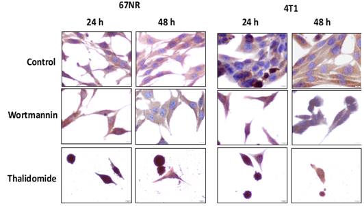
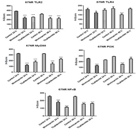
The immunoreactivities of TLR2 were positively strong in control groups of 67NR cell line Figure 1. In Wortmannin groups it was seen that TLR2 immunostaining were moderate Figure 1 and also compared to control groups their H-Score values were statistically significant (p<0.001) Figure 6. In Thalidomide 24-h treated group the TLR2 immunostaining was moderate/strong while in Thalidomide 48-h treated group it was moderate Figure 1 and also compared to control groups their H-Score values were statistically significant (p<0.001))Figure 6, Table 1.
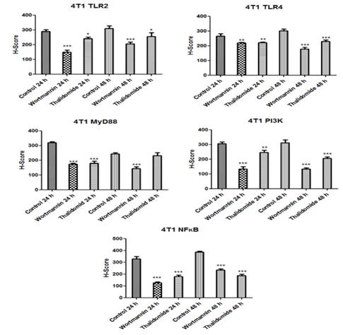
For 4T1 cell line the 24-h and 48-h control group immunoreactivities of TLR2 were moderate/strong and strong respectively Figure 1. For the wortmannin treated groups, their TLR2 immunoreactivities were detected as mild for 24-h and mil/moderate for 48-h while the immunoreactivities of Thalidomide treated groups were seen as moderate for 4T1 cell line Figure 1. The H-Score values were compared, and it was found that the wortmannin treated groups were statistically significant compared to the control group (p<0.001) Figure 7 and Thalidomide treated groups were also statistically significant (p<0.05) Figure 7, Table 2.
Figure 1
|
|
|
Figure 1 The immunohistochemistry micrographs
of TLR2. Original Magnification: X400. Scale Bar: 10 μm |
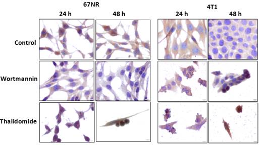
The TLR4 immunostainings were detected as moderate in control groups of 67NR cell line Figure 2. The TLR4 immunoreactivity of 24-h Wortmannin treated group was seen as mild/moderate and 48-h Wortmannin treated group was detected as mild while the Thalidomide treated groups were observed as moderate for 67NR cell line Figure 2. The H-Score values of 24-h Wortmannin treated group was statistically significant compared to the 24-h control group (p<0.05) and also 48-h Wortmannin treated group was statistically significant compared to the 48-h control group (p<0.001) Figure 6, Table 1. The Thalidomide treated groups of TLR4 H-Score values of 67NR cell line, there were not statistically significance compared to the control groups (p>0.05) Figure 6, Table 1.
For 4T1 cell line, the TLR4 immunoreactivity of 24-h control group was moderate/strong and 48-h control group was strong Figure 2. While the TLR4 immunoreactivities of Thalidomide treated groups were seen as moderate, the 24-h Wortmannin treated group was moderate and 48-h Wortmannin treated group was mild/moderate Figure 2. The H-Score values of TLR4 24-h Wortmannin and Thalidomide treated groups were statistically significant (p<0.01), and 48-h Wortmannin and Thalidomide treated groups also statistically significant (p<0.001) compared to the control groups of 4T1 cell line Table 2.
Figure 2
|
|
|
Figure 2 The immunohistochemistry micrographs
of TLR-4. Original Magnification: X400. Scale Bar: 10 μm |
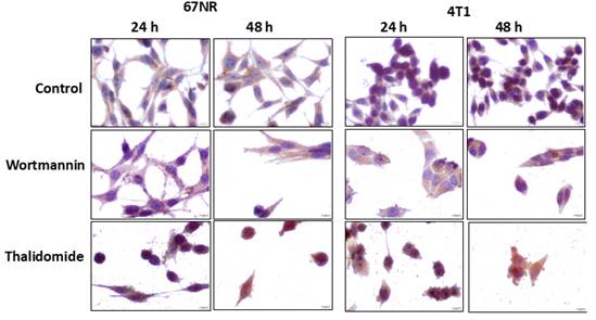
The immunoreactivities of MyD88 were found as strong in control groups of 67NR cell line Figure 3. The Wortmannin treated groups were detected as mild, the 24-h Thalidomide treated group was mild/moderate and 48-h Thalidomide treated group was moderate Figure 3. The H-Score values of MyD88 for 67NR cell line, it was seen that the treated groups were statistically significant compared to the control groups (p<0.001) Figure 6, Table 1.
For the 4T1 cell line, the immunoreactivities of MyD88 in control groups were detected as strong and moderate, respectively Figure 3. The MyD88 immunoreactivities of 24-h Wortmannin and 24-h Thalidomide treated groups was observed mild/moderate Figure 3. The immunoreactivity of MyD88 was found as mild for 48-h Wortmannin and as moderate for 48-h Thalidomide treated groups Figure 3. The H-Score values of 24-h Wortmannin, 24-h Thalidomide and 48-h Wortmannin treated groups were statistically significant compared to the control group (p<0.001) Figure 7, Table 2 while the H-Score values of 48-h Thalidomide treated groups was not statistically significant (p>0.05) Figure 7, Table 2.
Figure 3
|
|
|
Figure 3 The immunohistochemistry micrographs
of MyD88. Original Magnification: X400. Scale Bar: 10 μm |
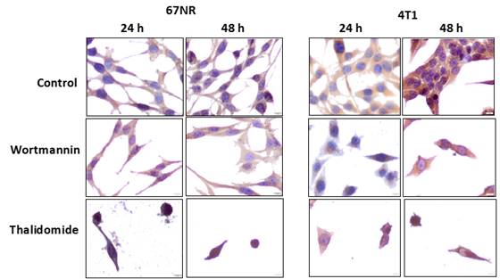
In the control groups of 67NR cell line, PI3K immunoreactivities were found as moderate in 24-h group and moderate/strong in 48-h group Figure 4. In the 24-h Wortmannin treated group, the PI3K immunoreactivity was detected as mild and in the 48-h Wortmannin treated group it was mild/moderate while PI3K immunoreactivity of 24-h and 48-h treated Thalidomide groups of 67NR cell line were moderate Figure 4. The H-Score values of PI3K of Wortmannin treated groups were statistically significant compared to the control groups of 67NR cell line (p<0.001) Figure 6, Table 1. In the 67NR cell line, the H-Score values of PI3K of 48-h Thalidomide treated group was statistically significant (p<0.001) however the 48-h Thalidomide treated group was not different (p>0.05) compared to the control groups Figure 6, Table 1.
The control groups of 4T1 cell line, the PI3K immunoreactivities were detected as moderate/strong Figure 4. In the Wortmannin treated groups the PI3K immunoreactivities were mild and in the Wortmannin treated groups they were moderate for 4T1 cell line Figure 4. The H-Score values of PI3K in Wortmannin treated groups were statically significant (p<0.001) and also in 24-h Thalidomide (p<0.01) and 48-h Thalidomide (p<0.001) treated groups were statically significant compared to the control groups for 4T1 cell line Figure 7, Table 2.
Figure 4
|
|
|
Figure 4 The immunohistochemistry micrographs
of PI3K. Original Magnification: X400. Scale Bar: 10 μm |
The NFκB immunoreactivities of 67NR cell line was moderate in control groups Figure 5. The immunostainings of NFκB in the Wortmannin treated groups and 48-h Thalidomide treated group were detected as mild/moderate while 24-h Thalidomide treated group was found as mild Figure 5. The H-Score values of NFκB in treatment groups were statically significant compared to the control group for 67NR cell line (p<0.001) Figure 6, Table 1.
For the 4T1 cell line the immunoreactivities of NFκB were seen as strong and very strong in control groups, respectively Figure 5. In the 24-h Wortmannin treated group, the NFκB immunoreactivity was detected as mild and in the 48-h Wortmannin treated group it was moderate Figure 5. The NFκB immunoreactivities of both 24-h and 48-h Thalidomide treated groups were seen as mild/moderate Figure 5. The H-Score values of NFκB in treatment groups were statically significant compared to the control groups for 4T1 cell line (p<0.001) Figure 7, Table 2.
Figure 5
|
|
|
Figure 5 The immunohistochemistry micrographs
of NFκB. Original Magnification: X400. Scale Bar: 10 μm |
The all proteins, which are evaluated in this study, showed decreased H-Score values. The H-Score values of all groups for all antibodies were given in Table 1 and Table 2.
Table 1
|
Table 1 The H-score values of each antibody of each group for 67NR cell line |
|||||
|
TLR2 |
TLR4 |
MyD88 |
NFκB |
PI3K |
|
|
Control 24 h |
383,33 ± 5,8 |
209 ± 12,8 |
322,33 ± 7,1 |
255,67 ± 8,6 |
253,67 ± 7,5 |
|
Wortmannin 24 h |
236,67 ± 23,6 |
171,67 ± 12,6 |
122,00 ± 12,1 |
180,00 ± 16 |
132,00 ± 8,9 |
|
Thalidomide 24 h |
256,67 ± 5,8 |
202,67 ± 14,5 |
178,67 ± 20 |
114,67 ± 6,1 |
231,67 ± 3,1 |
|
Control 48 h |
318,67 ± 12,9 |
218 ± 19,3 |
321,33 ± 4,5 |
253 ± 7 |
290,67 ± 4,0 |
|
Wortmannin 48 h |
227,33 ± 24,9 |
152 ± 10,6 |
145,67 ± 25,9 |
157,67 ± 6,7 |
156,33 ± 5,5 |
|
Thalidomide 48 h |
223,67 ± 24,6 |
215,67 ± 9 |
227,67 ± 13,6 |
163,33 ± 7,1 |
249,7 ± 18,2 |
|
Data is
expressed as means ± SD and were compared statistically |
|||||
Table 2
|
Table 2 The H-score values of each antibody of each group for 4T1 cell line |
|||||
|
TLR2 |
TLR4 |
MyD88 |
NFκB |
PI3K |
|
|
Control 24 h |
288,67 ± 12,1 |
265,33 ± 15,0 |
319,67 ± 6,1 |
330 ± 18,3 |
305,3 ± 12,9 |
|
Wortmannin 24 h |
149 ± 13,5 |
219,33 ± 4,5 |
174 ± 4,6 |
126,67 ± 8 |
133,7 ± 15,2 |
|
Thalidomide 24 h |
241,33 ± 9,0 |
221,67 ± 3,8 |
179,67 ± 13,6 |
178 ± 13,9 |
246 ± 14,9 |
|
Control 48 h |
310 ± 18,0 |
301,67 ± 10,8 |
243,67 ± 7,1 |
386 ± 8,7 |
311,7 ± 20,2 |
|
Wortmannin 48 h |
205 ± 13,2 |
177 ± 12,1 |
143,67 ± 11,9 |
233 ± 11,8 |
133,33 ± 7,0 |
|
Thalidomide 48 h |
255,33 ± 26,1 |
228,67 ± 10,3 |
230,67 ± 20,1 |
187,67 ± 12,1 |
205,33 ± 9,6 |
|
Data is expressed as means ± SD and were compared statistically |
|||||
Figure 6
|
|
|
Figure 6 H-Score values of each protein in 67NR
non-metastatic breast cancer cell line. (*p <0.05, **p <0.01, ***p
<0.001) |
Figure 7
|
|
|
Figure 7 H-Score values of each protein
in 4T1 metastatic breast cancer cell line. (*p <0.05, **p <0.01, ***p <0.001) |
4. DISCUSSION
Breast cancer has the highest incidence and mortality rate among women worldwide Jemal et al. (2011). Although it has been reported that breast cancer-related death rates have decreased in developed and high-income countries in recent years, the incidence has increased or remained stable. Although increasing incidence and mortality rates are alarming, rapid advances occur in health sciences in terms of cancer treatment DeSantis et al. (1978). The novel approach in this issue is to determine the molecular mechanisms in the cell signaling pathways that play a role in the progression of breast cancer. Oncogenic mutations targeting proteins in signaling pathways eliminate control of cell replication and/or survival. Oncoproteins play an important role in tumor development, invasion, and metastasis process by causing significant changes in the behavior of many cells regulatory signaling pathways Robert (2015).
TLRs have various functions in regulating cell homeostasis, survival, and cell death. It is known that TLR signaling pathway activation leads to the production of biological factors that create and activate inflammatory responses in the acquired immune system. TLRs can regulate cell proliferation and vitality by increasing tissue repair processes and inflammatory responses and create an environment for tumor cells to escape the immune response Basith et al. (2012).
Various mechanisms have been suggested for how TLRs organize the inflammatory response and carcinogenesis. The first of these is the anti-apoptotic effect of NFκB activated by the TLR signaling pathway. Secondly, TLRs demonstrate that oxidative DNA damage occurs, inducing tissue repair responses Rakoff-Nahoum and Medzhitov (2009) and by taking role in the molecular mechanisms that enable angiogenesis to occur with VEGF secreted by tumor cells, immune cells and cancer-related fibroblasts Carmeliet (2005). The relationship between breast cancer and TLRs is reported that TLR2 and TLR4 play an important role in breast cancer migration, invasion and also, they have angiogenic potential Bhatelia et al. (2014).
TLR2 signalling affects a dual-acting complex and dynamic process expressed by antigen presenting cells that enhances immunity against the tumor, and also increases tumor resistance and persistence Kim and Karin (2011). Clinical studies are still being conducted with TLR2 ligands for a wide variety of cancer types. While TLR agonists developed and used in these studies are expected to kill tumor cells by activating the host immune system, reducing tumor progression and metastasis, it is also stated that TLR2 agonists stimulate tumor growth Kim and Karin (2011).
In a study reported by Xie et al., it was found that invasive MDA-MB-231 cells showed that TLR2 expression was 10.5 times higher compared to less invasive MCF-7 cells. Also, TLR2 activation has been reported to cause increased levels of NFκB and increased IL-6, TGF-β, VEGF and MMP9 levels Xie et al. (2009).
TLR4 expression has been reported to be highly increased in cancer cells and immune cells in the tumor microenvironment. TLR4 has significant effects on the presentation of antigens in cancer cells as a result of chemotherapy and radiotherapy. TLR4 polymorphisms can affect the susceptibility of individuals to breast cancer development or recurrence. It is thought that TLR4 targeting in breast cancer cells has a role in reducing the metastatic potential of cancer cells Kim and Karin (2011). The expression of TLR4 in the invasive breast cancer cell line MDA-MB-231 cell line was very high compared to other TLRs, and silencing of TLR4 was found to inhibit cell proliferation and survival. In addition, it has been also reported inhibition or silencing of TLR4 has the potential to block breast tumor growth Yang et al. (2010).
In this study, it was determined that the intensity of TLR2 immunoreactivity was higher in the non-metastatic 67NR breast cancer cell line than the metastatic 4T1 breast cancer cell line. In 4T1 breast cancer cell line, it was determined that immunoreactivity of TLR4 was significantly higher. In the study, TLR2 immunoreactivity, which was observed as high in both breast cancer cell lines, it is thought to be effective on cancer cell proliferation and differentiation. When these findings are evaluated together, it is thought that TLRs are observed to be increased in breast cancer cells and TLR4 may play an important role as a trigger molecule in the emergence of invasive properties in cancer cells and initiation of the metastatic process.
MyD88 is an adapter molecule that plays an active role in all TLR signaling pathways. After it was revealed that TLRs play a role in cell proliferation and tissue repair, the role of MyD88 in cancer has been investigated. In a study conducted with knock-out mice, it was reported that TLR4 triggers colon cancer development and the number and size of tumors decreased in the absence of TLRs or MyD88 Fukata et al. (2007). In this study, MyD88 immunoreactivity was found to be high in breast cancer cell lines. It is thought that the increase of MyD88 in the carcinogenesis process provides suitable conditions for tumor development by increasing the release of tumor-supporting chemokines and cytokines. Considering the resistance-forming role of TLRs and assuming that they induce inflammatory processes, it has been concluded that MyD88 and therefore TLRs are necessary molecules in carcinogenesis and metastasis processes.
In immune cells, TLR expression is generally inhibited by the long-term activation of NFκB through negative feedback Biswas and Lopez-Collazo (2009). NFκB activation by the TLR signaling pathway causes the activation increased anti-apoptotic proteins and decreased pro-apoptotic proteins Nakanishi and Toi (2005). It has been reported that NFκB enables tumor cells to increase their lifespan, develop resistance to cancer drugs and allow the tumor to progress Nakanishi and Toi (2005). On the contrary, in another report of triple negative breast cancer cell lines (MDA-MB-231, MDA-MB-468, SUM-149 and SUM-159) it was reported especially TLR2, TLR3 and TLR4 accompanied by the continuous activation of NFκB and it has been shown to be selectively expressed. This suggests that TLRs are more effective in regulating NFκB activation Mehmeti et al. (2015).
In this study, high NFκB immunoreactivity was observed in the 4T1 cells and moderate in the 67NR cells. Looking at these results, it is seen that NFκB could play an effective role in the proliferation and survival of cancer cells. In 4T1 breast cancer cells, it was thought that having more NFκB than 67NR has an effect on cell proliferation, angiogenesis, and anti-apoptotic processes, which are the steps of invasion and metastasis processes.
Direct proof of PI3K participation in TLR signalling was first performed by Arbibe et al., via site-directed mutagenesis of specific tyrosine residues found in the cytosolic portion of TLR2. As a result of this mutagenesis, the ability of p85 to relate to TLR2 and the ability of TLR2 to transcribe NFκB was eliminated Arbibe et al. (2000). The cytosolic part of the TLR2 receptor contains an evolutionarily conserved binding site with PI3K binding Laird et al. (2009). It has also been shown that PI3K has a direct link with TLRs and adapter molecules of TLRs (such as MyD88). Activation of AKT occurs by PI3K with TLR stimulation Ruse and Knaus (2006).
In this study, positive immunoreactivity of PI3K, a signalling pathway that functions importantly by activating mTOR and AKT molecules in cell growth, proliferation, and protein synthesis, was observed. PI3K immunoreactivity in 67NR and 4T1 breast cancer cell lines was moderate and moderate/strong, respectively. PI3K immunoreactivities were found to be correlated with TLRs, and this correlation suggested that the PI3K molecule was also activated by TLRs.
Wortmannin is a steroid metabolite obtained from Penicillium funiculosum that inhibits PI3K molecules. Wortmannin was shown to induce MCF-7 cell death by causing chromatin condensation, nucleus fragmentation, reactive oxygen species formation and membrane changes, which are typical features of apoptosis Akter et al. (2012). Wortmannin has also been shown by Yun et al., to have proliferation-inhibiting and apoptosis-inducing effects on the MCF-7 human breast cancer cell line, dose and time depending on manner Yun et al. (2012). It has been also reported that this effect of Wortmannin is achieved by down-regulation of NFκB protein expression and by altering the regulation of the PI3K/AKT signaling pathway Yun et al. (2012).
With the immunohistochemical analysis, a significant decrease in cell number and a statistically significant decrease in the immunoreactivity intensities of TLR2, TLR4, MyD88, NFκB and PI3K were observed in breast cancer cell lines treated with Wortmannin compared to control groups. When the Wortmannin treatments were compared depending on the time, no significant difference was observed between immunoreactivities. Compared to the control group breast cancer cell lines showing positive immunoreactivity of PI3K, which plays an effective role in the proliferation and survival of cancer cells, a statistically significant decrease in the intensity of PI3K immunoreactivity in the Wortmannin treated groups confirmed that the drug is effective on this pathway. With this study, it was determined that the immunoreactivities of molecules that are effective in TLR signalling pathways were decreased in both breast cancer cell lines with Wortmannin treatments compared to the control groups. It is thought that Wortmannin can be used in the treatment of breast cancer as a therapeutic agent.
Thalidomide (immunoprin), which is an old antiemetic agent and known for its teratogenic properties, is currently used as an anti-angiogenic and immunomodulator. Initial results with thalidomide analogues show promise for future clinical studies by providing the desired anti-cancer activity and reducing its side effects Cook and Figg (2010). In the immunohistochemical study, in the 67NR and 4T1 breast cancer cell lines, when the control groups and the Thalidomide treated groups were compared, a significant decrease in cell number and a significant decrease in the immunoreactivity intensities of TLR2, TLR4, MyD88, NFκB and PI3K were observed. When Thalidomide treatments were compared depending on time, no significant difference was observed between immunoreactivities. It is thought that Thalidomide can be used in breast cancer as a therapeutic agent.
When the findings were evaluated by immunohistochemistry, Wortmannin administration was observed to be more effective on TLR2, TLR4, MyD88 and PI3K signal molecules while Thalidomide administration was significantly more effective on NFκB.
When activation of TLR2 and TLR4 molecules, the intracellular activator molecule MyD88 signaling begins. Later, NFκB, which takes part in the last step of the TLR signaling pathway and initiates the transcription of genes necessary for cell proliferation and survival, is activated and increased angiogenesis, chemoresistance, apoptosis inhibition, change of inflammatory response and triggering of the metastatic process begins. At the same time, activation of MyD88 activates the AKT pathway through PI3K, resulting in inhibition of apoptosis, cell proliferation, and increased survival. As a result of this study, it was thought that in vivo inhibition or silencing of TLR2 and TLR4 could be a potential treatment method to prevent tumor growth and the results put forward for targeting TLR2 and TLR4 may be related between breast cancer oncogenesis and future perspectives.
ACKNOWLEDGMENTS
This work was supported by Research Project Coordination Unit of the Manisa Celal Bayar University, project number 2014-171.
REFERENCES
Agnese DM, Calvano JE, Hahm S, Coyle SM, Corbett SA, Calvano SE, Lowry SF, (2002). Human Toll-like receptor 4 mutations but not CD14 polypmorphism are associated with an increased risk of gramnegative infections. The Journal of Infectious Diseases, 186(10),1522-152. https://doi.org/10.1086/344893
Akter R, Hossain MZ, Kleve MG, Gealt MA. (2012). Wortmannin induces MCF-7 breast cancer cell death via the apoptotic pathway, involving chromatin condensation, generation of reactive oxygen species, and membrane blebbing. 13(4),103-13. https://doi.org/10.2147/BCTT.S31712
Arbibe L, Mira JP, Teusch N, Kline L, Guha M, Mackman N, et al. (2000). Toll-like receptor 2-mediated NF-kappa B activation requires a Rac1-dependent pathway. https://doi.org/10.1038/82797
Basith S, Manavalan B, Yoo TH, Kim SG, Choi S. (2012). Roles of toll-like receptors in cancer : a double-edged sword for defense and offense. Journal of Occupational Medicine and Toxicology. 35(8),1297-316. https://doi.org/10.1007/s12272-012-0802-7
Becker S. (2015). A historic and scientific review of breast cancer : The next global healthcare challenge. International Journal of Gynaecology and Obstetrics. https://doi.org/10.1016/j.ijgo.2015.03.015
Bhatelia K, Singh K, Singh R. (2014). TLRs : linking inflammation and breast cancer. Cellular Signalling. 26(11), 2350-7. https://doi.org/10.1016/j.cellsig.2014.07.035
Biswas SK, Lopez-Collazo E. (2009). Endotoxin tolerance : new mechanisms, molecules and clinical significance. Trends in Immunology. 30,475-87. https://doi.org/10.1016/j.it.2009.07.009
Carmeliet P. (2005). VEGF as à key mediator of angiogenesis in cancer. Oncology. 69(3),4-10. https://doi.org/10.1159/000088478
Cook KM, Figg WD. (2010). Angiogenesis inhibitors : current strategies and futurs prospects. A Caner Journal for Clinicians, 60(4),222-43. https://doi.org/10.3322/caac.20075
DeSantis CE, Bray F, Ferlay J, Lortet-Tieulent J, Anderson BO, Jemal A. Dexter DL, Kowalski HM, Blazar BA, Fligiel Z, Vogel R, (1978). Heterogeneity of tumor cells from a single mouse mammary tumor. National Libarry of Medicine, 38(10), 3174-3181. https://pubmed.ncbi.nlm.nih.gov/210930/
Friis S, Kesminiene A, Espina C, Auvinen A, Straif K, Schüz J. (2015). European Code against Cancer 4th Edition : Medical exposures, including hormone therapy, and cancer. Cancer Epidemiology, 107-19. https://doi.org/10.1016/j.canep.2015.08.003
Fukata M, Chen A, Vamadevan AS, Cohen J, Breglio K, Krishnareddy S et al. (2007). Toll-like receptor-4 promotes the development of colitis-associated colorectal tumors. Gastroenterology. 133(6),1869-81. https://doi.org/10.1053/j.gastro.2007.09.008
Hirsch FR, Varella-Garcia M, Bunn PA Jr, Di Maria MV, Veve R, Bremmes RM, et al., (2003). Epidermal growth factor receptor in non-small-cell lung carcinomas : Correlation between gene copy number and protein expression and impact on prognosis. Journal of Clinical Oncology, 21(20), 3798-807. https://doi.org/10.1200/JCO.2003.11.069
Jemal A, Bray F, Center MM, Ferlay J, Ward E, Forman D. (2011). Global cancer statistics. A Cancer Journal for Clinicians, 61(2),69-90. https://doi.org/10.3322/caac.20107
John T, Liu G, Tsao MS. (2009). Overview of molecular testing in non-small-cell lung cancer : Mutational analysis, gene copy number, protein expression and other biomarkers of EGFR for the prediction of response to tyrosine kinase inhibitors. Oncogene, 14-23. https://doi.org/10.1038/onc.2009.197
Kim S, and Karin M. (2011). Role of TLR2-dependent inflammation in metastatic progression. Ann. The New York Academy of Science, 191-206. https://doi.org/10.1111/j.1749-6632.2010.05882.x
Laird MH, Rhee SH, Perkins DJ, Medvedev AE, Piao W, Fenton MJ, et al. (2009). TLR4/MyD88/PI3K interactions regulate TLR4 signaling. Journal of Leukocyte Blology, 85,966-77. https://doi.org/10.1189/jlb.1208763
Mehmeti M, Allaoui R, Bergenfelz C, Saal LH, Ethier SP, Johansson ME, Jirström K, Leandersson K. (2015). Expression of functional toll like receptor 4 in estrogen receptor/progesterone receptor-negative breast cancer. Breast Cancer Research, 17(1),130. https://doi.org/10.1186/s13058-015-0640-x
Nakanishi C, Toi M. (2005). Nuclear factor-κB inhibitors as sensitizers to anticancer drugs. Nature Reviews Cancer. 5(4), 297-309. https://doi.org/10.1038/nrc1588
Ozgul M, Turkoz Uluer E, Tanriover G, Inan S. (2016). Evaluation of the Apoptotic Effects of Wortmannin and Thalidomide on Breast Cancer Cell Lines. Turkish Journal of Biochemistry.
Rakoff-Nahoum S, Medzhitov R. (2007). Regulation of spontaneous intestinal tumorigenesis through the adaptor protein MyD88. Science, 124-7. https://doi.org/10.1126/science.1140488
Rakoff-Nahoum S, Medzhitov R. (2009). Toll-like receptors and cancer. Nature Reviews Cancer, 9,57-63. https://doi.org/10.1038/nrc2541
Robert J. (2015). Textbook of Cell Signaling in Cancer. Springer. https://doi.org/10.1007/978-3-319-14340-8
Ruse M, Knaus UG. (2006). New players in TLR-mediated innate immunity : PI3K and small Rho GTPases. 34,33-48. https://doi.org/10.1385/IR:34:1:33
Thike AA, Chng MJ, Fook-Chong S, Tan PH. (2001). Immunohistochemical expression of hormone receptors in invasive breast carcinoma : correlation of results of H-score with pathological parameters. Pathology, 33(1), 21-5. https://doi.org/10.1080/00313020120034858
Troutman TD, Bazan JF, Pasare C. (2012). Toll-like receptors, signaling adapters and regulation of the pro-inflammatory response by PI3K. 11(19),3559-67. https://doi.org/10.4161/cc.21572
Underhill D M, Ozinsky A, (2002). Toll-like receptors : key mediators of microbe detection. Current Opinion in Immunology, 14 (1), 103- 110. https://doi.org/10.1016/S0952-7915(01)00304-1
Xie W, Wang Y, Huang Y, Yang H, Wang J, Hu Z. (2009). Toll-like receptor 2 mediates invasion via activating NF-kappaB in MDA-MB-231 breast cancer cells. Biochemical and Biophysical Research Communications, 1027-32. https://doi.org/10.1016/j.bbrc.2009.01.009
Yang H, Zhou H, Feng P, Zhou X, Wen H, Xie X, et al. (2010). Reduced expression of Toll-like receptor 4 inhibits human breast cancer cells proliferation and inflammatory cytokines secretion. Journal of Experimental and Clinical Cancer research. https://doi.org/10.1186/1756-9966-29-92
Yuan TL, Cantley LC. (2008). PI3K pathway alterations in cancer : variations on a theme. Oncogene. 5497-5510. https://doi.org/10.1038/onc.2008.245
Yun J, Lv YG, Yao
Q, Wang L, Li YP, Yi J. (2012). Wortmannin inhibits
proliferation and induces apoptosis of MCF-7 breast cancer cells. 33(4),367-9. https://article.imrpress.com/journal/EJGO/33/4/pii/1631087281572-1634603826/367-369.pdf
 This work is licensed under a: Creative Commons Attribution 4.0 International License
© Granthaalayah 2014-2022. All Rights Reserved.