0BINTRODUCING GAP IN HAIR FOLLICLE ELECTROMAGNETISM AS PROPOSED MECHANISM FOR THE PRESENCE OF BIPOLAR ELECTRICAL CHARGES INHERENT IN THE HUMAN HAIR SHAFTAbrahám A. Embí BS 1 1B1 13442 SW 102 Lane, Miami,Florida 33186, United States |
|
||
|
|
|||
|
Received 5 September2021 Accepted 16 September2021 Published 30 September2021 Corresponding Author Abrahám
A. Embí BS, embi21@att.net DOI 10.29121/granthaalayah.v9.i9.2021.4260 Funding:
This
research received no specific grant from any funding agency in the public,
commercial, or not-for-profit sectors. Copyright:
© 2021
The Author(s). This is an open access article distributed under the terms of
the Creative Commons Attribution License, which permits unrestricted use, distribution,
and reproduction in any medium, provided the original author and source are
credited.
|
ABSTRACT |
|
|
|
The
human hair consists of a follicle anchored in the skin and a protruding
shaft, it has also been described as a miniorgan, having its own cell
divisions, metabolism, and known to undergo aging stages; eventually reaching
a point where the old hair sheds and a new hair growing cycle begins from the
same follicular tissue. Using sophisticated magnetometers, magnetic field
emitted by direct current (DC) in human hair follicles were detected and
introduced in 1980. Most recently in 2015, a tabletop optical microscopy
method was developed and published in 2016, thus allowing for the detection
of hair follicles and shaft magnetic fields. Qualitative images are presented
where the bipolar electrical property of the shaft is documented. This
finding was inferred since blood tissue carries a negative charge, thus
repelled by an equal charge; experiments support a positive (+) field as
triggering coagulation. The shaft is repeatedly shown in experiments to
express a contralateral positive side inducing clots. Fibrin formation is
also documented by images showing intricate networks indicative of blood
coagulation. In conclusion, the genesis of hair shafts bipolarity is shown
resulting from a “gap” in the follicle electromagnetic fields inhibiting
energy from fully engulfing the shaft. |
|
||
|
Keywords: Potassium
Blocking Rejection, Hair Follicle, Bipolar Hair Shaft, Hair as dc Battery,
Hemocoagulation, Shepherds Hook Genesis, Tissue DC Currents, Hair Follicle
Gap, Hair External Electromagnetism Definition of Terms DC = Direct Current. K3Fe
= Acronym for Potassium Ferricyanide K₃[Fe(CN)₆]. EMR =
Electromagnetic Radiation 1. INTRODUCTION The human hair consists of a follicle
anchored in the skin and a protruding shaft, it has also been described as a miniorgan,
having its own cell divisions, metabolism, and known to undergo aging stages
(Schneider et al. (2009)); eventually
reaching a point where the old hair sheds and a new hair growing cycle begins
from the same follicular tissue. Using sophisticated magnetometers, magnetic
field emitted by direct current (DC) in human hair follicles were detected
and introduced in 1980 (Cohen et al. (1980)). Most recently
in 2015, a tabletop optical microscopy method was developed and published in
2016, thus allowing for the detection of hair follicles and shaft magnetic
fields (Scherlag et al. (2016)). When a hair
shaft is in contact with fresh blood tissue on a slide, an interesting
finding occurs, which is one side inducing blood coagulation and the opposite side
inhibiting of coagulation. |
|
||
2. MATERIALS AND METHODS
2.1. Materials
Potassium Ferricyanide K₃[Fe(CN)₆].
25x75x1 mm glass slides
Fresh human blood smear
Freshly in toto plucked scalp human hair
Demineralized bottle water
Celestron Video Microscope Model # 44348
MacBook Pro Apple computer with Photo Application software.
2.2. Methods
1) A fresh human blood smear obtained. Solution prepared by diluting approximately 2 milligrams of (K3Fe) diluted in one drop of demineralized water was added to the center of a wet smear*.
Note: [1] * A finger stick allowed for the milking of two drops of blood, then placed on a clean 25x75x1 mm glass slide. The mechanical smear was done as per published instructions from the USA center for disease control. There is a time window of approximately 60±20 seconds for a complete preservation of in vivo properties of the blood tissue. For details link to: (https://doi.org/10.5281/zenodo.3472760)
2) One freshly tweezers plucked scalp hair was carefully placed in approximately the area where the of the liquid K3Fe had displaced the blood tissue.
3) The preparation allowed evaporating, Images recorded and stored for analysis.
3. RESULTS AND DISCUSSIONS
Prior research by this author showed the follicle and shaft magnetic fields properties on a glass slide (in the absence of blood) with the addition of only liquid K3Fe, as shown in images showing the hair shaft unilateral presence of electromagnetic radiation (EMR) (Embi (2018)) (Figure 2). In this manuscript, qualitative images are presented where not only the bipolar electrical property of the shaft is documented; but also showing EMR originating from the hair follicle routed only to one side of the shaft, a gap in the hair follicle’s EMR continuity accounting for the shaft’s bipolarity (+ −) (Figure 2, Figure 3).
Potassium as Immunosuppressing Agent
The rejection
of several exogenous materials, such as a hair follicle, keratin flakes and
small iron filings amongst others by a fresh human blood smear had been
described, Figure 7 ( Embi (2018)).
In this
manuscript, the addition of liquid K3Fe to a fresh human
blood smear inhibited rejection of an in
toto hair (follicle and shaft). This inhibition allowed for images such as
in (Figure 2, Figure 3, Figure 4, Figure 5). The question arises: What is the
mechanism whereby addition of liquid K3Fe to a fresh
blood smear inhibits fresh blood from rejecting a hair follicle?
Perhaps some
elucidation could be explained by findings where a link between tumor-induced
immune suppression by the Potassium ion (K+) exists (Vodnala et al. (2019)).
K3Fe Total Absorption of
Incoming EMRs
Hair Shaft
Polarity and Blood Coagulation
Liquid K3Fe
allowed for the display of hair follicle and shaft EMR as shown (Figure 2, Figure 4). A second important observation is the findings or previous papers correlating a
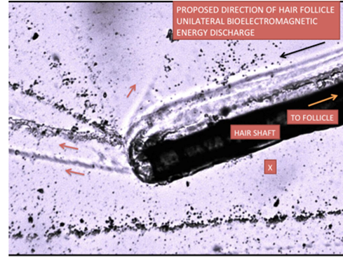
positive charge with blood coagulation. Early in 2018, an image was recorded
showing what appeared to be a one-sided electrical discharge in a human hair
shaft. (Figure 1). In 2018 there was not enough published
experimental data found by this author to support a hair shaft bipolarity
finding.
The question
arose: How could a keratin surrounded filamentous structure express opposite
sides charges?
Prior
Publication Hinting at Bipolarity
Potassium
Ferrocyanide Spatially Detachment of Shaft Exo-Cuticles
A paper published
in 2016, showed that when a human hair shaft was sandwiched between two glass
slides and covered by Potassium Ferrocyanide (Embi (2016)), post evaporation, microscopy images showed
an unexplained phenomenon, being the spatially separated images of hair
exocuticles. In other words, there was a need of a microscope depth of field
adjustment to bring the cuticles layers in focus as shown in Figure 6 below.
Evidence shows that the addition of one drop of liquid K3Fe onto a fresh blood smear inhibited rejection of a foreing material, namely a plucked human hair. This inhibition, allowed for the identification of the hair EMRs. This is supported by a property of K3Fe being the “Full absorption od incoming EMRs” ( Figgis et al. (1969), Baranov et al. (2015))
The hair unique
electromagnetic radiation pattern is shown partially engulfing the follicle.
There is a “gap” shown in the images that does not allow the magnetic signal
from traveling to one side of the hair shaft. The Hair Shaft Inherent
Unilateral Divergent Charges (+ −) are displayed.
The Dual Consequences of
Follicle’s EMR Gap
First) The abrupt
interruption of EMRs in one side of the hair follicle, accounts for the hair
shaft’s one-sided EMR activity triggering positive charges.
Second) The genesis of a
visual display resembling a “Shepherd Hook” pattern of the human hair EMR
emissions.
|
|
|
Figure 1 Hair shaft sanwiched between glass slides and
covered by drops potassium Ferrocyanide in solution. Showing shaft unilateral
discharge of elecromagnetic energy. Black Arrow: Direction of forces- X:
Shaft side void of energy. |
Image reproduced from: Cite This Article: Abraham A. Embi Bs. (2018). “THE SHEPHERDS HOOK PHENOMENON PATTERN OF HAIR ROOTS A DEMONSTRATION OF COMPARATIVE BIOLECTROMAGNETISM BETWEEN HUMAN HAIRS AND MOUSE WHISKERS BY MEANS OF THE PHOTOELECTRIC EFFECT.” International Journal of Research - Granthaalayah, 6(7), 317-326. https://doi.org/10.29121/granthaalayah.v6.i7.2018.1312.
|
|
|
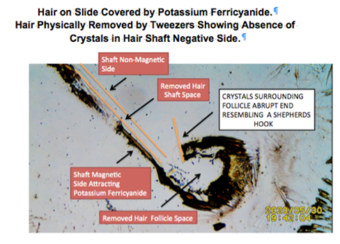
Figure 2 Image of hair imprint surrounded by human blood
tissue mixed with potassium Ferricyanide crystals. Notice the EMR unable to
fully reach one side of the hair shaft (orange line). |
Image reproduced from: Embi, AA (2021). Some curious findings
hair follicles bioelectromagnetic radiation expressed as light displacing
matter in its path and the contralateral emission of magnetic fields found in
the hair shaft. International Journal of Research - GRANTHAALAYAH, 9(7),
334. doi: 10.29121/granthaalayah.v9.i7.2021.4114
Another Hair Placed on Human Blood Smear Plus Liquid Potassium Ferricyanide Drops
|
|
|
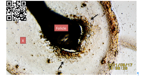
Figure 3 N=2 Hair on fresh human blood
smear, post addition of K3Fe allowing for this demonstration.
Black Arrow: Pointing at mix of K3Fe
crystals and coagulated blood surrounding follicle prior removal via
tweezers. |
For further
details link to: https://youtu.be/LLz43yAbpg0….or Scan QR Code in
left upper corner of image
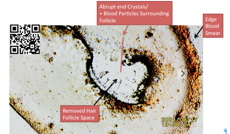
Same Hair as in Figure 3 Above. Hair Removed via Tweezers Showing Follicle’s Imprint Delineating EMRs Abruptly Ending Prior to Surrounding the Follicle. Red line indicating, “gap” void of EMRs.
|
|
|
Figure 4 image introduced in this
manuscript showing Hair EMR outline after hair removed. Black Arrows:
Pointing at mix of crystals and coagulated blood plus edge of blood smear.
The abrupt end of EMRs circumventing the follicle is shown. |
For details
link to: https://youtu.be/LLz43yAbpg0….or Scan QR Code in left upper corner of image.
Amplified Image of Figure 4 above
|
|
|
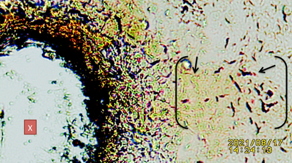
Figure 5 Amplified
image of Fig 4, showing X= Some blood tissue trapped under hair follicle.
Brackets= Showing mix of blood tissue (RBCs) and fibrin. |
Image
published in 2016 Hinting at Bipolarity of Hair Shaft Exocuticles
|
|
|
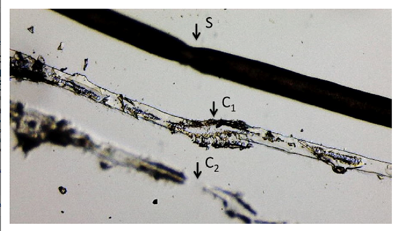
Figure 6 Microphotograph
of hair in contact with Prussian Blue (PBS) between two glass slides (SDW)
after evaporation, showing: S= Shifting shaft showing damage caused by
tweezers, C1 = In focus damaged anterior level cuticles, C2 = Out of focus
damaged posterior level cuticles (out of focus). X4 Magnification. |
Image reproduced from:
Embi AA. Adhesion Failure of External Hair Cuticles
Caused by Prussian Blue: Possible Electrochemical Roles of Sulfur and Cystine.
J Nat Sci, 2(6):e194, 2016.
Image of Fresh
Human Blood Smear Repulsing Hair Follicle
|
|
|
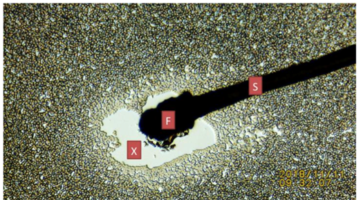
Figure 7 Image
depicting fresh human blood smear rejecting
hair follicle. F= Follicle. X= Empty space due to repulsive
phenomenon. |
Image
reproduced from: Cite This Article: Abraham A. Embi Bs. (2018).
“BIOMAGNETISM AS FACTOR IN RED BLOOD CELLS DEFORMATION.” International
Journal of Research - Granthaalayah, 6(12), 46-57.
https://doi.org/10.29121/granthaalayah.v6.i12.2018.1245.
Image published in 2018 of Hair Shaft on Fresh Blood Smear
Showing Effect of Positive Pole on Coagulation
|
|
|
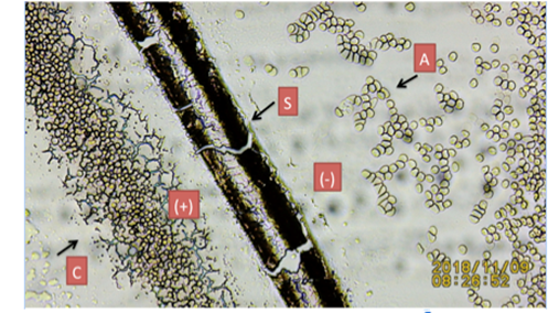
Figure 8 Microphotopraph depicts detached hair shaft outline. |
A= Non-coagulated
blood S= Hair shaft C= Coagulated blood.
(-)= Negative pole RBC repulsion. (+)= Positive pole attracting RBCs (coagulation). Reproduced from:
Abraham A. Embi Bs. (2018).
“HAIR AND BLOOD ENDOGENOUS LOW LEVEL BIOMAGNETIC FIELDS CROSS-TALK EFFECTS ON
FIBRIN INHIBITION AND ROULEAU FORMATION.” International Journal of Research
- Granthaalayah, 6(11), 200-208. https://doi.org/10.29121/granthaalayah.v6.i11.2018.1118.
4. SUPPLEMENTAL INFORMATION
Upon re-visiting my files,
found some video-recordings where the hair was physically removed from the
glass smear- Since the hair had been covered by Potassium Ferricyanide (K3Fe)
in solution; and K3Fe has the property of full absorption of
incoming electromagnetic radiation (EMR), the hair outer layers EMR are shown
as K3Fe crystals. These images are introduced for the first time in
this manuscript- Notice the consistent narrowing between the distal follicle
and the bulb-
|
|
|
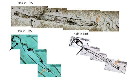
Figure 9 Showing sequential images from video-recordings
outlining the human hair external
electromagnetic radiation. Black Arrows: Notice the narrowing shown
between the distal follicle and the bulb also showing a gap in energy
continuity. This gap is theorized to induce the bipolar nature of the shaft
(+-). TIBS= Temporary In Vivo Blood Smear. Orange Arrows: Pointing at area
where shaft exits towards skin. |
|
|
|
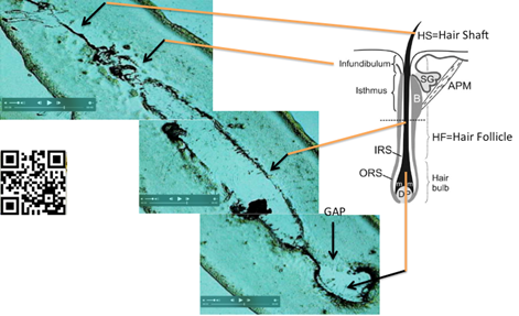
Figure 10 Cut and Paste frames to illustrate the human scalp hair magnetic
imprint post hair removal from slide. Scalp hair in fresh blood smear mixed
with liquid Potassium Ferricyanide. After drying, outer layers of hair are
delineated showing hair anatomical areas. Human hair magnetic Imprint on
glass slide. Drawing on right side of figure reproduced as described in the
copyright fair use doctrine. |
Credit of drawing given to:
Pisal Rishikaysh et al. DOI: doi:10.3390/ijms15011647
For video detais link to:: https://youtu.be/LLz43yAbpg0 Or scan QR Code in left side of Figure 4.
REFERENCES
Abraham A. Embi Bs. (2018). “BIOMAGNETISM AS FACTOR IN RED BLOOD CELLS DEFORMATION.” International Journal of Research - Granthaalayah, 6(12), 46-57. Retrieved from https://doi.org/10.29121/granthaalayah.v6.i12.2018.1245.
Abraham A. Embi Bs. (2018). “THE HUMAN HAIR FOLLICLE PULSATING BIOMAGNETIC FIELD REACH AS MEASURED BY CRYSTALS ACCRETION.” International Journal of Research - Granthaalayah, 6(7), 290-299. Retrieved from https://doi.org/10.5281/zenodo.1341349.
Abraham A. Embi Bs. (2018). “THE SHEPHERDS HOOK PHENOMENON PATTERN OF HAIR ROOTS A DEMONSTRATION OF COMPARATIVE BIOLECTROMAGNETISM BETWEEN HUMAN HAIRS AND MOUSE WHISKERS BY MEANS OF THE PHOTOELECTRIC EFFECT.” International Journal of Research - Granthaalayah, 6(7), 317-326. Retrieved from https://doi.org/10.29121/granthaalayah.v6.i7.2018.1312.
B. N. Figgis, Malcolm Gerloch, Ronald Mason, and Ronald Sydney (1969) Nyholm the crystallography and paramagnetic anisotropy of potassium ferricyanide. https://doi.org/10.1098/rspa.1969.0031 Retrieved from DOI: https://doi.org/10.1098/rspa.1969.0031
Cohen, D., Palti, Y., Bn, C. & Sj, S. (1980). Magnetic Fields Produced By Steady Currents In The Body. Proc. Natl. Acad. Sci 77(3), 1447–1451. Retrieved from https://doi.org/10.1073/pnas.77.3.1447
Embi AA (2016). Adhesion Failure of External Hair Cuticles Caused by Prussian Blue: Possible Electrochemical Roles of Sulfur and Cystine. J Nat Sci, 2(6):e194.
D. G. Baranov, J. H. Edgar, Tim Hoffman, Nabil Bassim, Joshua D. Caldwell (2015). Perfect interferenceless absorption at infrared frequencies by a van der Waals crystal. DOI: 10.1103/PhysRevB.92.201405 Retrieved from DOI: https://doi.org/10.1103/PhysRevB.92.201405
Scherlag, B.J., Sahoo, K., Embi, A.A (2016). A Novel and Simplified Method for Imaging the Electromagnetic Energy in Plant and Animal Tissue. Journal of Nanoscience and Nanoengineering 2(1): 6-9.
Schneider MR, Schmidt-Ullrich R, Paus R (2009). The hair follicle as a dynamic miniorgan. Curr Biol. Feb 10;19(3): R132-42. Retrieved from doi: 10.1016/j.cub.2008.12.005. Retrieved from https://doi.org/10.1016/j.cub.2008.12.005
Vodnala S.K., Eil R., Kishton R.J., Sukumar M., Yamamoto T.N., Ha N.H., Lee P.H., Shin M., Patel S.J., Yu Z., Palmer D.C., Kruhlak M.J., Liu X., Locasale J.W., Huang J., Roychoudhuri R., Finkel T., Klebanoff C.A., Restifo N.P (2019). T cell stemness and dysfunction in tumors are triggered by a common mechanism.Science. Mar 29. Retrieved from https://doi.org/10.1126/science.aau0135
 This work is licensed under a: Creative Commons Attribution 4.0 International License
© Granthaalayah 2014-2021. All Rights Reserved.