
PULMONARY THROMBO-EMBOLISM: DIFFICULTIES ENCOUNTERED IN ESTABLISHING CAUSAL RELATIONSHIP AND EXCLUDING THERAPEUTIC MISMANAGEMENT-A CASE REPORT
Abstract
Pulmonary embolism designates blockage of pulmonary arteries by substances moved from elsewhere in the body through the circulation. The commonest form is thromboembolism. The presentation of pulmonary thromboembolism (PTE) is extremely variable from being completely asymptomatic to sudden cardiovascular collapse and death. Shortness of breath, chest pain and haemoptysis are other common presentations. PTE is not a frequently encountered entity in routine autopsy practice. Out of all cases detected at autopsy, approximately fifty percent are not clinically diagnosed or even suspected, making it one of the top pathological entities clinically underdiagnosed or misdiagnosed. The autopsy, therefore remains the gold standard of post-mortem identification of PTE. Autopsy also allows to establish or exclude a clinico-pathologic co-relation between the clinical diagnosis and actual cause established post-mortem. Legal issues will arise as to whether a particular traumatic event has a causal relationship with the cause of death as PTE. Establishment of the cause of death (COD) as PTE, timing of the thrombotic event in relation to an ante-mortem trauma and exclusion of medical negligence on the basis of misdiagnosis and missed diagnosis are some important medico-legal issues encountered by the forensic pathologist. The objective of this case presentation is to elaborate practical difficulties encountered by the pathologist in addressing above medico-legal issues. The death of a 46-year-old male bike-rider from PTE, who died three days after being discharged from the hospital on the 26th day following a traffic accident resulting in multiple rib fractures and severe abdominal trauma which warranted splenectomy is discussed here.
Keywords
Pulmonary Thromboembolism, Deep Vein Thrombosis, Trauma, Traffic Accidents, Special Stains
INTRODUCTION
Embolism means obstruction of a blood vessel by a foreign substance or a blood clot which travels through the circulation. When embolism occurs due to a dislodged blood clot, it is termed thrombo-embolism which is by far the commonest form of embolism. (Kumar & Kaca, 2018; Saukko & Kb, 2015) Other forms of emboli include fat, bone-marrow, parasites, air, amniotic fluid, septic clumps of organisms, tumours and foreign material such as talc or even bullets. One common target organ to suffer thromboembolism is the lungs. This entity is termed pulmonary thromboembolism and results when one or both pulmonary arteries or their branches are plugged and blocked by thrombi. Most cases of PTE are resultant to deep venous thrombosis (DVT) of lower limbs and less commonly of other sites such as pelvic veins. Depending on the amount of involvement of the lungs, numbers and sizes of the thrombi, recurrent nature of the condition as well as the presence of underlying lung and heart diseases, the outcome and the presentation of PTE will vary from an asymptomatic or mild presentation to sudden death following cardiovascular collapse. (Kumar et al., 2018) Common signs and symptoms may include sudden onset shortness of breath which usually worsens with exertion, chest pain similar to an acute ischaemic event of the heart, cough with tinges of blood, rapid and irregular heart-beat, dizziness, profuse sweating, mild fever, cyanotic extremities and features suggestive of deep venous thrombosis of legs such as pain, tenderness and swelling of the calves. Most of these symptoms and signs are vague and non-specific and may even mislead to a clinical diagnosis of an acute ischaemic cardiac event. Several risk factors have been recognized including medical conditions and treatments to some such conditions as cardiovascular disease, certain types of cancers, surgery, clotting disorders leading to hypercoagulability, renal disorders, prolonged bed-riddenness, long distance air travel, smoking, obesity, oestrogen supplementation, pregnancy and trauma. (Kumar et al., 2018; Saukko et al., 2015) When a person succumbs to PTE following a significant traumatic event with or without surgery, the question arises as to the level of contribution of the trauma (which could be an accident, suicidal event or a homicidal attempt) to result in pulmonary thrombo-embolism. Another issue the forensic pathologist conducting the autopsy will have to address is the contribution of surgery towards PTE. In both conditions, the ageing of the thrombus at its origin/site of attachment to the vessel wall will have to be estimated-an entity which cannot be accomplished with mathematical accuracy in most instances. PTE, as mentioned earlier, is one of the commonest entities which is clinically underestimated and looked over for more common conditions such as ischaemic heart disease. (Both, Bruni, & Herath, 2018; R, 2013) The missed diagnosis or misdiagnosis will pose a question as to whether the clinician has payed reasonable degree of care and skill in the context of duty of care towards the patient sometimes leading to litigations of possible medical negligence. (R, 2013) The forensic pathologist who conducted the autopsy may invariably be called upon as an expert witness in such litigations. Further, the autopsy may also be helpful in establishing a clinico-pathologic correlation between the tentative ante-mortem diagnosis and the definitive cause of death arrived at the autopsy. If special dissections are not employed with or without special investigations and due caution is not exercised during the autopsy, certain marginal cases of PTE may be missed even at the post-mortem examination. (Both et al., 2018)
CASE PRESENTATION
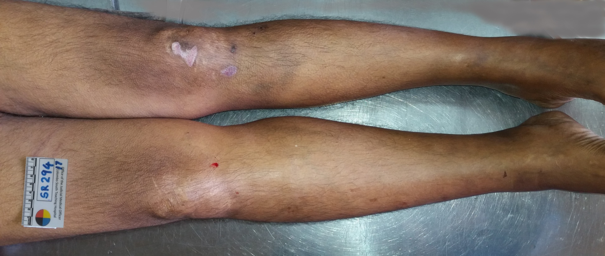
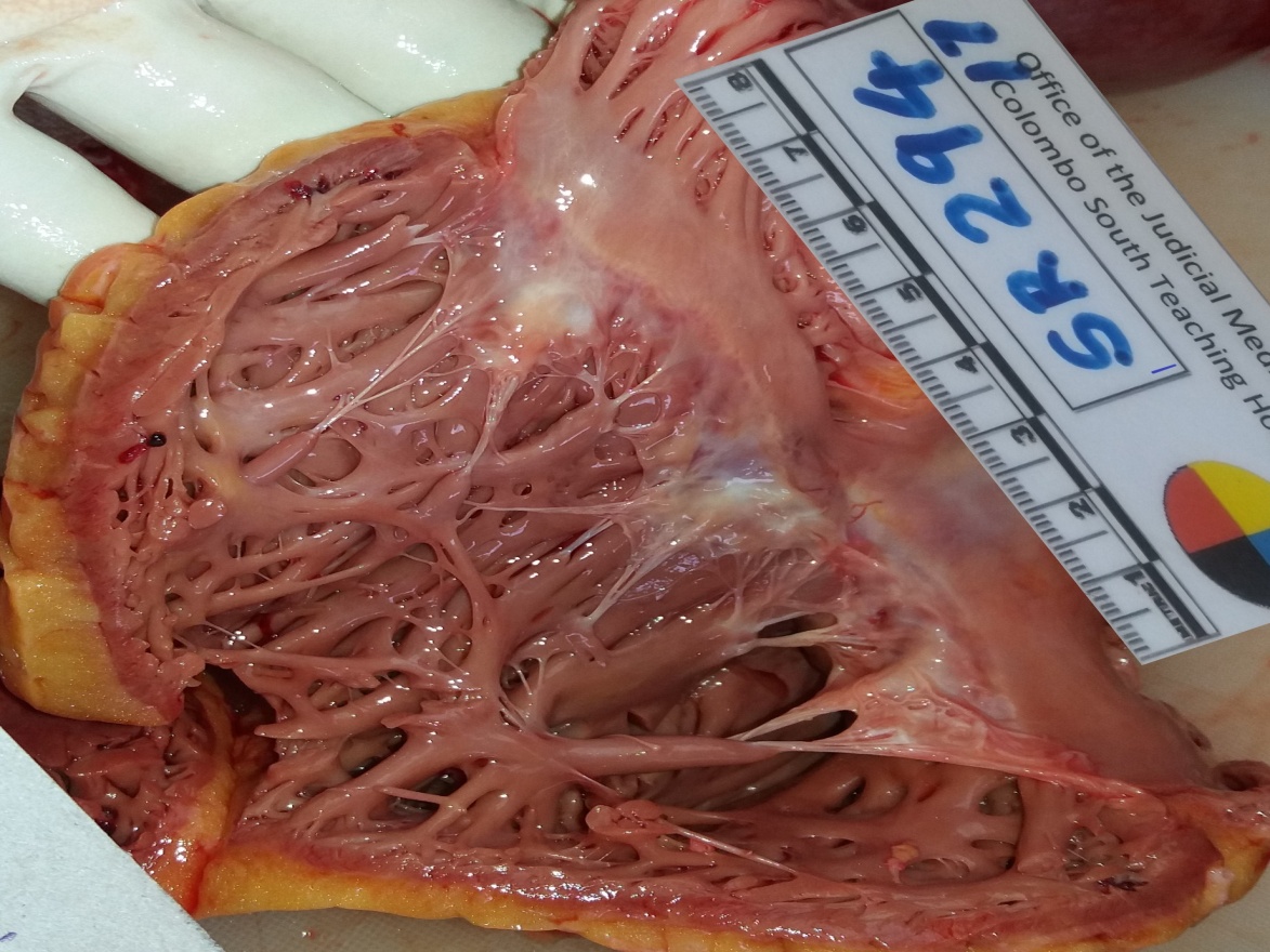
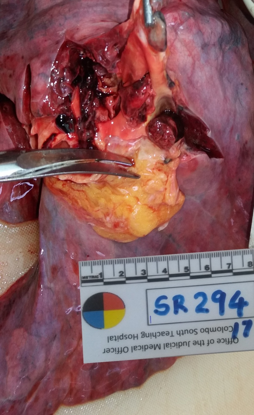
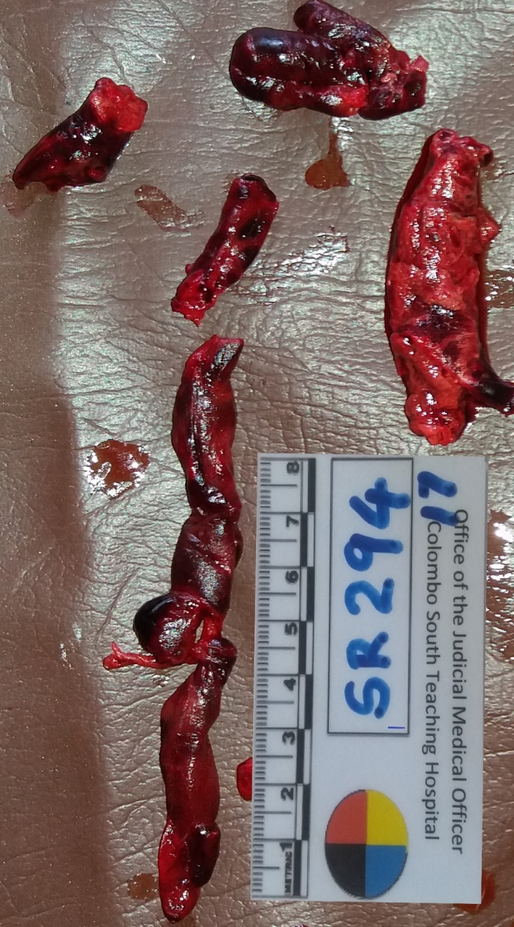
A 46-year-old male was admitted to the Emergency Treatment Unit of a tertiary care hospital following sudden onset of difficulty in breathing, irregular pulse, sweating and cold, clammy, cyanotic extremities. Despite initial resuscitative measures, he died within the first hour of admission. He had been discharged from the same hospital three days back following a 23-day-long stay in the same hospital and an uneventful recovery from an emergency laparotomy and splenectomy due to splenic rupture/laceration he had sustained in a road traffic accident 26 days prior to his death. While he was riding his motor-bike, he fell off the bike when trying to avoid a stray dog on the road and hit the left side of his chest and anterior abdomen against a pile of stones by the side of the road. He had been admitted to hospital with chest pain exacerbating with respiratory attempts. Chest x-ray showed fractured ribs from the 5th to the 9th on the left side. Abdominal ultra sound scan showed haemoperitoneum with lacerated spleen. Exploratory laparotomy excluded damage to other intra-abdominal organs. Splenectomy was performed and the patient was kept in the ICU for thirteen days. During this time, he had developed post-surgical pneumonia probably due to lung contusions which had been treated in the ICU set-up with IV antibiotics. Pneumococcal, meningococcal and HIB vaccines were given. He was haemodynamically stable, though not fully mobile due to pain. Three days after the discharge, he had suddenly developed severe shortness of breath while he was in the bath-room. He was again brought to the same hospital where he died within the first hour of admission. Due to the unusual nature of the death, an inquest had been conducted and a judicial autopsy had been requested at the inquest. External examination at the autopsy revealed the swollen nature of the right lower limb specially below the knee joint, multiple healed abrasions on the knee joints and the feet and the midline laparotomy incision and the drain sites all of which were healing well without signs of infection. In the internal examination, the brain was unremarkable. Dilatation of the right ventricle with relative thinning out of the wall with a thickness of just 0.2 cm was noted in the heart. A large thrombo-embolus was found obstructing the right pulmonary artery and the lower part of the main pulmonary trunk. Few patchy consolidated areas in the lower lobes of both lungs were evident. Spleen was absent and the splenic bed was devoid of any infection or haemorrhage from surgical site. The inferior vena cava, hepatic and portal veins as well as the pelvic vessels were all devoid of thrombi. Dissection of the right calf for deep venous thrombosis demonstrated a deep-seated, relatively large, organized thrombus within the entire length of the popliteal vein. Histologically (when stained with Haematoxylin and Eosin) the affected areas of the lungs showed resolving pneumonia with alveolar wall infiltration with haemosiderin-laden macrophages and lymphocytes. The embolus obtained from the pulmonary vasculature showed lines of Zahn with alternate bands of pale and red areas consisting of fibrin and platelets together with degrading red blood cells. No blood samples had been taken for investigations during the initial period of resuscitation immediately after the admission to the emergency treatment unit as the initial management had been more focussed on achieving clinical stability of the patient during the relatively short period he survived after admission. Post mortem blood culture and toxicological analysis were unremarkable. The cause of death was coined according to the WHO guidelines as follows:
-
Pulmonary thrombo-embolism
-
Deep vein thrombosis of lower limb
-
Prolonged immobility, abdominal surgery and complications of road traffic trauma


DISCUSSION
Difficulties encountered by the pathologist in establishing PTE at autopsy are multiple. (Both et al., 2018) A pathologist performing an autopsy should have a high degree of suspicion to detect PTE and this suspicion should be formulated upon the case presentation as well as the background of the deceased, past medical and social history of the deceased. Without this high level of suspicion, the autopsy pathologist may be carried away by the ante-mortem clinical diagnosis and the in-situ dissection of the pulmonary trunk would not be performed or he may not tend to perform relevant postmortem imaging to detect PTE even when such facilities are available. (Sane & Zkma, 2013; Saukko et al., 2015) Post-mortem clots have to be excluded when arriving at a diagnosis of PTE. This could be done macroscopically and more reliably microscopically.
Post-mortem clots take the contour of the vessel in which it lies. The branching pattern is same or similar to the vessel where it was found. Ante-mortem thrombi are curled as they traverse through the circulation from its site of origin to where it was found. Post-mortem clots are gelatinous, rubbery and take the texture and appearance of ‘currant jelly on chicken fat’. (Both et al., 2018) On microscopic examination, ante-mortem thrombi may show distinct histological appearance including the so called lines of Zahn. (Kumar et al., 2018; Saukko et al., 2015) An ante-mortem thrombus is already dislodged from its origin and as such to establish the site of origin careful dissection of the deep veins of the suspected area (mostly lower limbs) would be needed. Though the dating of a thrombus could not be achieved with mathematical accuracy, an attempt with aid of histology is worth trying when establishing ‘the cause and the effect’ type of relationship is important for the criminal justice system.
Pulmonary thrombo-embolism has a very variable incidence according to autopsy data which ranges between 1% to 30% depending on the clinical condition. (Both et al., 2018; Kerkez et al., 2009) Minimal incidence is noted among the general hospitalized populations who die due to numerous other causes mostly not related to burns, trauma or fractures. Highest incidence is recorded among deaths associated with burns, trauma, fractures, disseminated infection, extensive surgery and pregnancy complications. (Amendola & Optd, 2019; Eom & Jysckhtk, 2014; Kumar et al., 2018; R, 2013; Sane et al., 2013; Saukko et al., 2015) Almost all (approximately 95%) pulmonary thrombo-emboli arise from the large deep veins of the lower limbs where the thrombus has propagated to involve the popliteal veins. (Eom et al., 2014) In this case under discussion too, the origin was from the deep veins of the right lower limb.
Some common risk factors include prolonged immobilization specially immobilization involving the lower limbs, surgery specially orthopaedic procedures involving the hip and knee, severe trauma including extensive burns, multiple fractures and crush syndrome, congestive cardiac failure, use of oral contraceptives containing high levels of oestrogens, advanced pregnancy, disseminated cancer and primary hyper coagulopathy disorders such as factor V Leiden mutation to activated protein C, prothrombin gene mutation, deficiencies of anti-thrombin I, II and III, protein C and S deficiencies etc. (, 2010; Gudipati & Fecvhsspknea, 2014; Mansueto & Cdcevfbgcrea, 2019; Starr & Szspsdeaabea, 2019) The deceased under discussion had met with a traffic accident, sustained multiple blunt trauma including several rib fractures and splenic lacerations and had undergone a major surgery and had been bed-ridden for few weeks following the scenario making him an ideal candidate for deep vein thrombosis and PTE.
The sequelae of DVT and PTE are diverse. Some authors claim that 60%-80% of cases are clinically silent due to several factors including the minute size of the emboli, their non-recurrence and the number of emboli being limited. Obstruction of small to medium sized pulmonary vasculature may lead to pulmonary infarctions and this represents around 10%-15% of the cases. Pulmonary infarctions are commoner in the presence of some degree of circulatory insufficiency. Acute right sided heart failure and cardiovascular collapse would lead to death in 05% of the cases, when more than 60% of the total pulmonary vasculature is blocked. (C, 2007; Starr et al., 2019) This has happened to the deceased due to the large size of the thrombus and its site of impact. ( (, 2010; C, 2007) In a minority of patients which accounts for around 03% of all cases, chronic recurrent showers of minute emboli may lead to the development of pulmonary hypertension and resultant right heart failure. (Kerkez et al., 2009; Rich, Mkha, & Trauma, 2004; Shonyela, Yang, Liu, & Jiao, 2015)
Determining the causal relationship between PTE and any speculated previous traumatic event (assault, accident or any other form of trauma or such similar event) is of considerable medico-legal significance. This may well be an exclusion; that PTE has arisen not subsequent to but prior to the event in question. The embolus may well be the most recent addition to an extending venous thrombus which is considerably old. Therefore, timing/dating should focus not only on the dislodged embolus but also the origin of the thrombus. As such, the thrombo-endothelial junction at the origin of the thrombus provides the most reliable data for ageing of DVT and PTE. (Dzidzava et al., 2021; Fineschi & Tenmpcri, 2009; Hope & Dbnwstslhbea, 2007; Mansueto et al., 2019) As such, the best approach is to examine the residual thrombus which is almost always found in the deep leg veins. The vein wall containing the origin of the thrombus is obtained as a segment of the thrombosed vein preferably with the surrounding muscles during the dissection for histological examination. Special staining techniques may be employed for dating of a thrombus and histological interpretation would need adequate experience and exposure as a histopathologist. PTAH (phosphotungstic acid-haematoxylin) stain will stain fibrin as purplish strands on the first day. Small masses and thicker strands or sheets may be visible by the fourth day. Evidence of endothelial proliferation would be most useful within the first week. Small buds begin to arise from the vessel wall by around the second day and proliferate over the first week. Collagen fibres do not tend to appear in the initial period and they may be visible only after five to ten days’ time. Fibroblasts may be seen as early as in the first two-three days though they usually tend to appear towards the end of the first week and reaching a maximum by the second or the third week. (Amendola et al., 2019; Mansueto et al., 2019) Sometimes they tend to increase in numbers until the fourth week. Haemosiderin-laiden macrophages are maximally seen around the third week. Elastic fibres usually do not appear before the fourth week. The current three-stage refinement of DVT grading scheme proposed by Fineschi et al (2009) is based on immunohistochemistry and Confocal Laser Scanning Microscopy (CLSM). (Fineschi et al., 2009) The authors claim that using immunohistochemistry and CLSM, only three chronological stages (described as phase I, II and III in the original research) can be distinguished based on the transformational histological features shown by the thrombus undergoing organization. Accordingly, after two months, the thrombus appears hyalinized with central sinuous cavities, indicative of a primary collagen composition produced by fibroblasts. Densely packed collagen fibers may contain sparse immune cells, looser tracts of collagen are associated with neovascularization. The cross-section of the thrombus shown in the fig. 3 is similar to this description (though it is limited to H&E staining alone) and as such will fall under phase III of Fineschi classification which confirms that it is older than two months. This clearly shows that the traumatic episode (traffic accident and the subsequent surgery) had occurred on a date much later than the origin of the onset of DVT.
CONCLUSION
Pulmonary thrombo-embolism in association with traumatic scenarios, surgeries, burns etc. poses several medico-legal issues to the autopsy pathologist some of which may be materially significant issues before courts of law. One such important issues is the dating of the process of thrombo embolism in relation to the traumatic event in question. Histopathological examination of the thrombo-endothelial junction with special stains and techniques would be of help in delineating the time of thrombus formation.
AUTHORS’ CONTRIBUTIONS
Both authors have contributed equally
CONSENT
Consent had been obtained from the senior next of kin of the deceased for displaying photographs though identity of the deceased could not be traced by the case history or photographs.
ETHICAL APPROVAL
Not relevant as the autopsy had been done under the authority of the inquirer in to sudden deaths as a part and parcel of a legal procedure.
COMPETING INTERESTS
No competing interests between authors
