Original Article
A computational Docking and Structure-Activity Relationship analysis of the Phytochemical Isorhamnetin as a potential inhibitor of 2vsm (Glycoprotein of Nipah virus)
|
Rajorshi Sen Gupta 1* 1 Department of Computer
Science and Applications, Malda College, Malda, India |
|
|
|
ABSTRACT |
||
|
In the world of zoonotic virus, Nipah virus is one of the severe pathogenic virus and very much responsible for neurological disorders and highly respiratory diseases with increasing mortality rates. To dominate this fatal virus specific antiviral therapeutic system is required, that’s why searching for the novel inhibitors are very much needed. This study represent a flavonoids, Isorhamnetin, a phytochemical of Acacia catechu wans deployed as an potent actsnhibitor against candidate Glycoprotein of Nipah virus (PBD:2vsm) via analysis of Molecular docking. Autodock software gave the result of molecular docking (dlg file) and that showed the binding affinity was -6.32 kcal/mol. This result indicate the protein interaction of binding site was stable. Structural demonstration of the isorhamnetin was shown by SwissAdme and according to that this phytochemical connect with 2vsm, the macromolecule with multiple hydrogen and hydrophobic bonds interactions with the residues including Asn557, Ser561, Lys560, Leu552, and Val555. This study also highlighted the Structure–activity relationship (SAR) analysis by giving importance of hydroxyl and methoxy groups in enhanced binding affinity and stability. According to findings it may suggest that as a inhibitor, isorhamnetin act as a promising character against Nipah virus contamination. Keywords: Isorhamnetin, Nipah Virus, 2VSM,
Molecular Docking, Flavonoidsm Antiviral,
Structure–Activity Relationship |
||
INTRODUCTION
Nipah virus is a
Fatal emerging pathogen which belongs to zoonotic diseases are a member of Paramyxoviridae virus family which is a significant global
threat regarding public health Chua et al. (2000), Ang et al. (2018). By contamination of this virus fatal
encephalitis and respiratory disorders may occurs in human and in some
outbreaks, mortality rates already exceeded 70% Ang et al. (2018), Lo and Rota (2008).
The virus
attachment is done with the viral attachment Glycoprotein (PDB:2vsm) and it
plays a significant role for recognize host cell and viral entry. This make it an attractive target for antiviral drug development Newman
and Cragg (2020).
Natural herbal
compounds, particularly secondary metabolites under the domain of flavonoids
gets its attention due to the antiviral and other safety properties Clayton
(2017).
The Isorhamnetin
is a secondary metabolites of Acacia catechu which is
a well known plant used as
medicinal plants in Indian traditional therapeutic system. This phytochemical
of Acacia catechu is a methylated flavonol and has the antiviral, antioxidant and
anti-inflammatory properties Calderón-Montaño
et al. (2011)
The aim of this
study is to reveal potential that inhibitors properties of Isorhamnetin against
the Glycoprotein. The study demonstrated binding interactions through molecular
docking and SAR analysis.
Materials and Methods
The crystal 3D structure of the Glycoprotein of Nipah virus (PDB ID: 2VSM) was searched out from the RCSB Protein Data Bank Database.
|
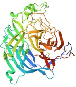
Figure 1
|
|
Figure 1 Nepha Virus Glycoprotein with Human Control Surface
Receptor Ephrinb2 |
2vsm the PDB
Number of glycoprotein of Nipah Virus. The structure
contains Chain A, which is virus content and Chain B which is human control
surface receptor ephrinB2 Xu et al. (2008). For that reason
the download PDB file was taken to Discovery Studio where Chain B was
eliminated. After that all the heteroatom and water was
removed by the protein structure and Kollman charges were added and saved the
file as PFBQT format.
Ligand Preparation
|
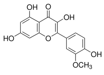
Figure 2
|
|
Figure 2 Chemical Structure
of Ligand Isorhamnetin |
The 3D sdf file of Isorhamnetin was obtained from PubChem. Then it
was taken to OpenBable to find out the PDB structure.
The ligand, Isorhamnetin was taken to Avogadro2 software for was
energy-minimized with MMFF94 force field and hydrogen atoms were added. Then
Gasteiger charges were assigned and converted it into PDBQT format.
Molecular Docking
Both
PDBQT file was taken to AutoDock 4.2 software. Lamarckian Genetic Algorithm was
applied. The grid box was centered around the active
binding pocket of 2VSM. AutoGrid and AutoDock was applied for molecular docking and the docking
log (.dlg) file was analyzed
to extract binding energies and interaction conformations.
|
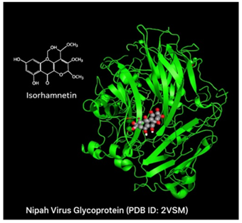
Figure 3
|
|
Figure 3 Complex Structure
After Molecular Docking |
Structure–Activity Relationship (SAR) Analysis
SAR analysis was
performed to evaluate the role of functional groups in ligand binding.
Structural features such as hydroxyl groups, methoxy substitution, and aromatic
rings were correlated with binding affinity.
|
Figure 4
|
|
Figure 4 Binding Pocket of
2vsm at Complex Structure of Isorhamnetin and 2vsm |
Results and Discussion (Docking and Structure–Activity Relationship)
The analysis of
Molecular Docking of 2bsm with Isorhamnetin 2VSM showed a binding energy of
−6.32 kcal/mol, that indicates a strong interaction and good binding
stability. The Isorhamnetin as a ligand was occupy the active binding site of
the glycoprotein efficiently which revealed it’s
potentiality as an antiviral inhibitor. The results of docking ( .dlg file) showed a strong binding energy, which was
suggested that the Isorhamnetin ligand was generated a stable complex within
the binding active site of the protein. The binding model demonstrated that
ligand is well accommodated in the 2vsm (receptor) cavity, adopting an position that maximizes intermolecular interactions.
Interaction Analysis of Molecular Docking
Detailed analysis
of the docked complex showed that ligand Isorhamnetin poses multiple hydrogen bonds with key polar
residues like Ser and Asn, which are vital for
anchoring the ligand Isorhamnetin within the binding pocket. These transactions
arise primarily from the hydroxyl (–OH) groups which are present on the
flavonoid scaffold, which act as both hydrogen bond donors and acceptors. Such
interactions significantly enhance binding specificity and stability.
In addition to
polar transaction, the ligand Isorhamnetin also engages in hydrophobic
relations with non-polar amino acid residues including Leu and Val, inside the
binding cavity. These transactions contribute to the stabilization of the
ligand Isorhamnetin through van der
Waals forces and hydrophobic packing. The presence of a methoxy (–OCH₃)
group in isorhamnetin increases its lipophilicity, thereby strengthening its
interaction with hydrophobic regions of the protein.
A vital feature
shown in the docking study is the presence of π–π stacking
interactions between the aromatic rings of ligand Isorhamnetin and aromatic
residues such as phenylalanine and tyrosine. The planar flavonoid backbone
enables effective overlap of π-electron clouds with these residues at an
optimal distance (~3.5 – 4.5 Å), causes to enhanced electronic stabilization.
These π–π transaction play a crucial role in reinforcing the
ligand–protein complex and improving binding affinity.
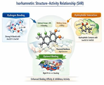
SAR (Structure–Activity Relationship) Interpretation
Key Interacting
Residues
Hydrogen Bonds:
Asn557, Ser561, Lys560
Hydrophobic
Interactions: Leu552, Val555
Van der Waals
interactions: Tyr581 and surrounding residues
The inhibitory
activity of ligand Isorhamnetin can be directly correlated with its structural
features:
Methoxy
Substitution (–OCH₃)
The methoxy group
helps to increased lipophilicity, facilitating better interaction with
hydrophobic amino acids such as Leu and Val. This modification also helps in
improving membrane permeability, an important pharmacokinetic property.
Hydroxyl Groups
(–OH)
The presence of
multiple hydroxyl groups secure the formation of good
hydrogen bonds with active site residues, thereby increasing binding affinity
and specificity.
Flavonoid
Aromatic Backbone
The rigid and
planar structure of the flavonoid core ensures optimal alignment within the
binding pocket. This structural rigidity minimizes conformational entropy loss
upon binding and promotes π–π stacking interactions with aromatic
residues, further stabilizing the complex.
Synergistic
Effect of Functional Groups
The combined
presence of hydrogen bonding groups, hydrophobic substituents, and aromatic
rings creates a multi-interaction binding mechanism, which significantly
enhances the overall inhibitory potential of the molecule.
Integrated
Insight
The docking
results and SAR analysis collectively demonstrate that ligand Isorhamnetin
exhibits a multi-modal interaction profile involving hydrogen bonding,
hydrophobic interactions, and π–π stacking. This combination of
interactions leads to a highly stable ligand–protein complex, which is
essential for effective inhibition of the Nipah virus glycoprotein, 2vsm.
|
Figure 5
|
|
Figure 5 Structure -Actibity Relationship (SAR) of Ligand Isorhamnetin |
The study
highlights that structural optimization of flavonoid derivatives, particularly
through modification of hydroxyl and methoxy groups, could further enhance
antiviral activity. Therefore, isorhamnetin serves as a promising lead compound
for the development of novel therapeutics targeting Nipah virus infection.
Conclusion
This study shows
that ligand Isorhamnetin exhibits strong binding affinity and stable
transactions with the Nipah virus glycoprotein (2VSM). The docking results,
supported by SAR analysis, indicate that functional groups such as hydroxyl and
methoxy moieties play a vital role in enhancing inhibitory activity. The
interaction with key residues such as Asn557, Ser561, and Lys560 highlights its
potential as a promising antiviral candidate.
Further studied Xu et al. (2008), including molecular dynamics simulations
and experimental validation, are required to confirm its therapeutic efficacy.
ACKNOWLEDGMENTS
None.
REFERENCES
Ang, B. S. P., Lim, T. C. C., and Wang, L. (2018). Nipah Virus Infection. Journal of Clinical Microbiology, 56(6), e01875-17. https://doi.org/10.1128/JCM.01875-1
Calderón-Montaño, J. M., Burgos-Morón, E., Pérez-Guerrero, C., and López-Lázaro, M. (2011). A Review on the Dietary Flavonoid Isorhamnetin: Biological Effects and Mechanisms of Action. Current Medicinal Chemistry, 18(32), 4922–4937.
Chua, K. B., Bellini, W. J., Rota, P. A., Harcourt, B. H., Tamin, A., Lam, S. K., Ksiazek, T. G., Rollin, P. E., Zaki, S. R., Shieh, W., Goldsmith, C. S., Gubler, D. J., Roehrig, J. T., Eaton, B., Gould, A. R., Olson, J., Field, H., Daniels, P., Ling, A. E., … Mahy, B. W. J. (2000). Nipah Virus: A Recently Emergent Deadly Paramyxovirus. Science, 288(5470), 1432–1435. https://doi.org/10.1126/science.288.5470.1432
Clayton, B. A. (2017). Nipah Virus: Transmission and Pathogenesis. Current Opinion in Virology, 22, 97–104.
Lo, M. K., and Rota, P. A. (2008). The Emergence of Nipah Virus. Journal of Virology, 82(12), 5399–5401.
Newman, D. J., and Cragg, G. M. (2020). Natural Products in Drug Discovery. Journal of Natural Products, 83(3), 770–803.
Xu, K., Rajashankar, K. R., Chan, Y. P., Himanen, J. P., Broder, C. C., and Nikolov, D. B. (2008). Host Cell Recognition by the Henipavirus Attachment Glycoprotein. Proceedings of the National Academy of Sciences, 105(29), 9953–9958.
 This work is licensed under a: Creative Commons Attribution 4.0 International License
© Granthaalayah 2014-2026. All Rights Reserved.